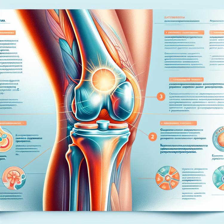
Schlatter’s disease: symptoms, diagnosis, and treatment methods
- Definition and Causes of Development of Scheuermann’s Disease
- Etiology of Schlatter’s disease
- Signs of the development of Schlatter’s disease
- Expert opinion on treatment methods for Schlatter’s disease
- Methods for diagnosing Schlatter’s disease
- Methods of treating Schlatter’s disease
- Prevention measures for Scheuermann’s disease
- Amazing features of Scheuermann’s disease
- FAQ
Definition and Causes of Development of Scheuermann’s Disease
Scheuermann’s disease, also known as osteochondropathy of the adolescent growth age, is a condition characterized by necrosis of the periosteum at the upper pole of the tibial tuberosity. The causes of this disease are not fully understood, but risk factors may include increased physical activity, injuries, genetic factors, and rapid growth development in adolescents.
It is believed that increased mechanical stress and microtraumas associated with growth changes and active participation in sports may contribute to the development of Scheuermann’s disease. Further research may shed light on the molecular mechanisms of this condition and help develop more effective treatment strategies.
Etiology of Schlatter’s disease
Schlatter’s disease, also known as knee osteochondrosis, typically develops in adolescents during periods of rapid growth. Its primary cause is considered to be overloading of the knee joint due to intense physical activity, sports training, or excessive use of the joint.
The main factors contributing to the development of Schlatter’s disease include structural features of the bones and joints, muscle and ligament fatigue, as well as lack of stretching and insufficient cushioning during prolonged physical exercises. The presence of damage or defects in the cartilage may also contribute to the development of this condition.
- Osgood-Schlatter disease is more common in adolescents during periods of active growth
- Intensive physical activity can contribute to the development of the disease
- The structural features of bones and joints may be a primary cause of the disease
- Lack of stretching and imperfect cushioning also play a role in the development of the disease
- Injuries or damage to cartilage can also lead to the development of Osgood-Schlatter disease
Signs of the development of Schlatter’s disease
Symptoms of the development of Osgood-Schlatter Disease typically include pain and swelling in the area above the knee. Patients may experience discomfort when moving the knee or during prolonged stress on it. It is important to note that symptoms may worsen during physical activity and improve at rest.
Other signs indicating the development of this condition include a feeling of instability in the knee, limited mobility, and possible swelling. In case of persistent symptoms or worsening condition, it is important to consult a doctor for diagnosis and appropriate treatment.
- Knee pain: patients may feel discomfort and tenderness when bearing weight on the knee.
- Swelling and edema: the area above the knee may be swollen due to inflammation and fluid accumulation.
- Sensation of instability: patients may feel that their knee is unstable and unable to bear their weight during movement.
- Limited mobility: the development of Schlatter’s disease may lead to difficulty in bending or straightening the knee.
- Worsening symptoms during physical activity: the symptoms of the condition may worsen with physical exertion and improve at rest.
Expert opinion on treatment methods for Schlatter’s disease
Expert opinions on the treatment methods for Schlatter’s Disease emphasize the importance of an individual approach to each patient. Specialists typically recommend a combined approach that includes conservative methods such as physical therapy, therapeutic exercise, and wearing specific types of protective braces.
Experts also note that in some cases, surgical intervention may be required, especially if conservative treatment does not lead to improvement in the patient’s condition. Surgical treatment methods may include the removal of major bone fragments or reducing irregularities in the cartilage surface to restore the normal structure of the knee joint.

Methods for diagnosing Schlatter’s disease
The diagnosis of Schlatter’s Disease includes a clinical examination of the patient to identify characteristic symptoms such as pain, swelling, limited mobility, and instability in the knee joint. Additionally, X-rays may be performed to determine the extent of changes in the bones and cartilage, as well as MRI or CT for a more detailed study of the joint structure.
Clinical data and examination results assist doctors in establishing an accurate diagnosis of Schlatter’s Disease and determining the most effective treatment plan for each patient. Early detection and diagnosis of this condition are essential for conducting effective therapy and preventing complications in the future.
- Clinical examination: The doctor conducts an examination to identify characteristic symptoms such as pain, swelling, and instability in the knee joint.
- X-ray: Used to assess structural changes in the bones and cartilage of the joint.
- Magnetic resonance imaging (MRI): Provides a more detailed image of the joint tissues for accurate disease diagnosis.
- Computerized tomography (CT): Allows for three-dimensional imaging of the joint to identify structural changes.
- General blood and urine tests: May help exclude other causes of symptoms and assess the overall condition of the patient’s body.
Methods of treating Schlatter’s disease
In cases of severe symptoms of Osgood-Schlatter disease or lack of results from conservative methods, surgical intervention may be necessary. Surgical methods include removing parts of the osteochondral fragment or correcting structural changes in the knee joint. Each patient requires an individual approach to treatment depending on the severity of the disease and the characteristics of its manifestation.
- Limitation of physical activity: It is important to reduce the load on the knee joint to decrease pressure and prevent further irritation.
- Use of anti-inflammatory medications: Medications in this group help reduce inflammation and alleviate pain in the affected joint area.
- Physical therapy: Exercises to strengthen the muscles around the knee and improve joint mobility contribute to the recovery of functionality and reduce the risk of new injuries.
- Surgical intervention: If conservative methods are ineffective, surgery on the knee joint may be required to correct structural changes.
- Individual approach: Treatment of Osgood-Schlatter disease requires consideration of the individual characteristics of the patient, the severity of the disease, and its manifestations to choose the optimal treatment strategy.
Prevention measures for Scheuermann’s disease
To prevent Osgood-Schlatter disease, it is important to pay attention to your health, strengthen the muscles around the knee joint, distribute physical activity correctly, and seek medical advice in a timely manner if pain or discomfort in the knee area occurs. Effective prevention helps reduce the risk of developing the condition and maintains joint health over the long term.
- Moderate physical activity: Regular moderate loads strengthen muscles and joints, reducing the risk of developing Osgood-Schlatter disease.
- Correct exercise technique: When engaging in sports or physical exercises, it’s important to follow the correct technique to avoid knee injuries.
- Preventing overexertion: It is necessary to avoid excessive strain on the knee, especially in adolescents or athletes, to prevent the development of the disease.
- Monitoring activity levels: It’s important to monitor the intensity of physical exercises and maintain a balance between activity and rest for knee health.
- Strengthening the muscles around the knee joint: Systematic exercises to strengthen muscles stabilize the joint and reduce the load on the knee, helping to prevent disease development.
Amazing features of Scheuermann’s disease
One of the interesting aspects of Schlatter’s disease is the possibility of self-regression with adherence to recommendations for limiting physical activity and rehabilitating the joint. This is due to the unique ability of the body to restore the structure of the joint with proper care and treatment. Understanding the characteristics of the development of this disease allows for the development of more effective methods of prevention and treatment.