Fibrous dysplasia: features of diagnosis and modern treatment methods
- Studying fibrous dysplasia: main aspects and characteristics
- Factors and mechanisms of the occurrence of fibrous dysplasia
- Visible manifestations of fibrous dysplasia
- The specialists’ perspective on the treatment methods for fibrous dysplasia
- Methods of detecting fibrous dysplasia
- Methods and strategies for treating fibrous dysplasia
- Measures to prevent fibrous dysplasia
- Unusual aspects of fibrous dysplasia
- FAQ
Studying fibrous dysplasia: main aspects and characteristics
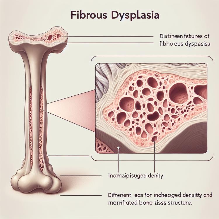
Fibrous dysplasia is a rare genetic disorder characterized by abnormal development of bone tissue, which leads to the formation of heterogeneous defects in bone structures. Pathological changes typical of fibrous dysplasia can occur in individual bones as well as in multiple bones simultaneously, complicating the diagnostic and treatment process.
Key aspects of fibrous dysplasia include changes in bone tissue structure, possible complications due to bone deformities, and the risk of fractures. Understanding the genetic and molecular mechanisms of this disease plays a crucial role in its diagnosis and therapy, with modern treatment methods aimed at improving the function of the affected bones and preventing complications associated with fibrous dysplasia.
Factors and mechanisms of the occurrence of fibrous dysplasia
Fibrous dysplasia is a rare genetic disorder characterized by a disruption in the process of bone tissue formation and development. The main cause of fibrous dysplasia is considered to be a somatic mutation in the genes responsible for regulating the differentiation of osteoblasts and osteoclasts. These mutations lead to the formation of dysplastic, poor-quality bone tissue cells, which causes disturbances in its structure and functioning. There are different subtypes of fibrous dysplasia, each characterized by unique genetic features and mechanisms of disease development.
- Genetic mutations: Somatic mutations in genes that control bone formation processes can trigger fibrous dysplasia.
- Cell differentiation disorders: Uncontrolled proliferation and development of dysplastic bone tissue cells contribute to the development of the disease.
- Impact of external factors: Exposure to toxic substances, radiation, or infections can cause changes in bone tissue, facilitating the development of fibrous dysplasia.
- Defects in the bone growth zone: Disruptions in the process of bone formation and growth at an early age can be a prerequisite for the emergence of fibrous dysplasia.
- Heredity: A family history of fibrous dysplasia increases the risk of developing the disease in descendants due to genetic predisposition.
Visible manifestations of fibrous dysplasia
Fibrous dysplasia can manifest with various symptoms, depending on the location of bone involvement and the extent of changes. One of the typical signs of fibrous dysplasia is tenderness or deformation of the bones, especially more frequently observed in the areas of long bones of the limbs. Patients may also experience restricted movements, pain during loading on the affected areas of the bones, as well as an increased risk of fractures due to the weakened structure of the bone tissue.
Other possible symptoms of fibrous dysplasia include external protruding bony formations (osteophytes), changes in the shape of the skull and face when the bones of the skull are affected, and enamel defects in cases of involvement of the jaw-face region. Early detection and differential diagnosis with other bone diseases allow timely treatment to be initiated and prevent the development of complications from fibrous dysplasia.
- Pain and deformation of bones: patients often experience pain and discover changes in the shape of bones, especially in the area of the limbs.
- Limited movements: abnormalities in the structure of bones can lead to restricted mobility in the affected joints and limbs.
- Increased risk of fractures: the weakened structure of bone tissue due to fibrous dysplasia increases the likelihood of fractures even with minor injuries.
- Protruding bone formations: the formation of osteophytes is observed, protruding on the surface of bones, which can cause discomfort and pain.
- Changes in the facial area: damage to the bones of the skull can lead to changes in the shape of the face, as well as problems with enamel and bite.
The specialists’ perspective on the treatment methods for fibrous dysplasia
Experts in the field of medicine believe that the treatment of fibrous dysplasia should be comprehensive and individualized, taking into account the characteristics of each patient and the nature of the disease. One of the main methods of treating fibrous dysplasia is surgical intervention aimed at correcting bone deformities, eliminating painful formations, and restoring the normal structure of bone tissue.
An important aspect of treating fibrous dysplasia is also medication therapy, aimed at reducing pain, maintaining bone and joint health, and strengthening bone tissue. Physiotherapy procedures and rehabilitation activities can also be included in the treatment plan to improve the functional condition of patients with fibrous dysplasia.

Methods of detecting fibrous dysplasia
The diagnosis of fibrous dysplasia includes various methods aimed at determining the characteristics of the affected bones. Radiological examination is one of the main diagnostic methods for fibrous dysplasia, allowing for the detection of changes in the structure of bone tissue and the identification of dysplastic elements. In addition, computed tomography (CT) and magnetic resonance imaging (MRI) can be used for a more detailed study of the affected areas and to assess the degree of changes.
Additional methods for diagnosing fibrous dysplasia include biochemical blood tests to assess the levels of certain markers of bone resorption and formation, as well as genetic testing to identify possible mutations responsible for the development of the disease. The comprehensive use of various diagnostic methods allows for an accurate diagnosis of fibrous dysplasia and the determination of the optimal treatment strategy for each patient.
- X-ray examination: One of the main diagnostic methods for fibrous dysplasia, allowing visualization of changes in bone structure and dysplastic elements.
- Computer tomography (CT): Used for detailed study of the affected areas and assessment of changes in bone tissue.
- Magnetic resonance imaging (MRI): Allows for detailed images of the structure of the affected bones, detecting possible disorders and anomalies.
- Biochemical blood analysis: Conducted to assess the level of certain markers of bone resorption and formation, which can aid in the diagnosis of fibrous dysplasia.
- Genetic testing: Used to identify mutations in genes responsible for the development of fibrous dysplasia, which helps confirm the diagnosis and determine the genetic basis of the disease.
Methods and strategies for treating fibrous dysplasia
An individual approach to treatment, taking into account the characteristics of the disease of each specific patient, contributes to improving their quality of life and preventing disease progression. Regular monitoring by specialists and a comprehensive approach to the therapy of fibrous dysplasia play an important role in achieving a favorable prognosis for the disease.
- Conservative therapy: Includes the use of anti-inflammatory and pain-relieving medications to reduce pain sensations and decrease inflammation.
- Physiotherapy: Physiotherapeutic procedures, such as ultrasound therapy, massage, and exercises, can help strengthen muscles and joints, improve blood circulation, and alleviate pain.
- Surgical intervention: In the case of significant bone deformities or fractures, surgical correction may be required to restore the structure and function of the affected areas.
- Rehabilitation: Rehabilitation activities, such as physical therapy sessions and reconstructive surgery, can help patients regain mobility and improve their quality of life.
- Individual approach: Each patient requires an individual approach to treatment, taking into account the specifics of their condition and physiological features to achieve optimal therapy results.
Measures to prevent fibrous dysplasia
Other preventive measures for fibrous dysplasia may include regular consultations with specialists (orthopedist, geneticist), screenings to detect early signs of the disease, as well as maintaining a healthy lifestyle that includes a balanced diet, physical activity, and avoiding harmful habits. All these measures will help reduce the risk of developing fibrous dysplasia and support overall health.
- Genetic counseling: Regular consultations with a geneticist can help identify hereditary predisposition to fibrous dysplasia.
- Family history: Knowing the family history of diseases can assist in early detection of risk factors and in the prevention of the disease in offspring.
- Regular examinations: Conducting regular examinations for early signs of fibrous dysplasia can help initiate treatment in time and prevent progression of the disease.
- Healthy lifestyle: Maintaining a healthy lifestyle, including a balanced diet, physical activity, and abstaining from harmful habits, contributes to overall body strengthening and may reduce the risk of various diseases, including fibrous dysplasia.
- Limiting risk factors: Avoiding harmful habits, managing weight, preventing injuries, and maintaining moderate levels of physical activity can help in the prevention of fibrous dysplasia.
Unusual aspects of fibrous dysplasia
Additionally, fibrous dysplasia can have various clinical manifestations, including pain in the affected bones, skeletal deformities, an increased risk of fractures, and other complications. Understanding the unusual aspects of this disease and its diverse manifestations helps specialists effectively diagnose and treat fibrous dysplasia, improving the prognosis and quality of life for patients suffering from this rare genetic disorder.