Fetal presentation: features, diagnosis, treatment.
- Understanding Brain Precedence: Key Aspects and Overview
- Pathophysiology of fetal cephalic presentation: influencing factors
- The concept of the outcomes of fetal head presentation
- Experts’ forecasts regarding the treatment of fetal cephalic presentation
- The main methods for diagnosing fetal head presentation.
- Methods for treating fetal head presentation
- Preventive measures to avoid fetal head presentation
- Amazing aspects of fetal cranial presentation
- FAQ
Understanding Brain Precedence: Key Aspects and Overview
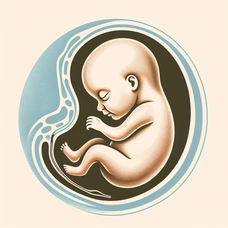
The head presentation of the fetus is a position in which the baby’s head is located at the upper part of the uterus before the onset of labor. This type of presentation occurs in most pregnant women and is considered the most favorable for normal natural childbirth. Key aspects of understanding head presentation include studying the anatomical features of the uterus, fetus, and amniotic sac, as well as the mechanisms of the fetus’s passage through the birth canal during the labor process. It is important to recognize the significance of regular pregnancy monitoring and timely identification of any deviations for successful management of pregnancy with head presentation.
Pathophysiology of fetal cephalic presentation: influencing factors
The pathophysiology of fetal cephalic presentation is associated with several important factors, including the size and shape of the maternal pelvis, the position and mechanics of fetal movement in the womb, fetal developmental anomalies, and the characteristics of the umbilical cord. Various influencing factors may contribute to the formation of cephalic presentation, including multiple pregnancies, fetal position, the presence of fetal anomalies, hydrops, or complications related to maternal health. Understanding these factors is important for assessing risks and choosing appropriate prevention and treatment methods for fetal cephalic presentation.
- Fetal anomalies: Some congenital anomalies, such as spinal development disorders, may contribute to cephalic presentation.
- Fetal position: An improper fetal position in the womb can increase the likelihood of cephalic presentation.
- Maternal pelvis: The size and shape of the mother’s pelvis can affect the possibility of the fetus passing through the birth canal normally.
- Multiple pregnancy: In the case of multiple pregnancies, the likelihood of cephalic presentation increases.
- Umbilical cord: Certain characteristics of the umbilical cord, such as its length or position, can also influence the development of cephalic presentation.
The concept of the outcomes of fetal head presentation
The concept of fetal vertex presentation outcomes represents an important medical aspect that reflects the prognosis and consequences of this condition for both the fetus and the mother. Outcomes of vertex presentation can vary from normal spontaneous delivery to the necessity of surgical intervention, such as cesarean section. It is important to consider and analyze the health indicators of the fetus and mother, possible complications during labor, as well as to take into account the individual factors of each clinical case when predicting the outcomes of fetal vertex presentation.
- Normal delivery: In the case of a normal and favorable delivery with the fetal head presentation, when no complications arise, labor proceeds without unexpected situations.
- Surgical intervention: Some cases of fetal head presentation may require surgical intervention, such as a cesarean section, for safe delivery and to minimize risks.
- Health indicators of the fetus and mother: Assessing the health status of both the fetus and the mother plays an important role in predicting outcomes of head presentation, as they determine possible risks and complications.
- Complications during labor: Some complications, such as dystocia and fetal injury during labor, may occur with fetal head presentation and affect outcomes.
- Individual factors of each clinical case: Considering the unique characteristics of each pregnancy and delivery, individual factors play an important role in predicting outcomes of fetal head presentation.
Experts’ forecasts regarding the treatment of fetal cephalic presentation
Experts’ opinions on the treatment of fetal occipital presentation reflect the importance of an individualized approach to each clinical case. Specialists emphasize the need for a thorough assessment of health indicators for both the fetus and the mother when making decisions about the treatment method. It is critically important to consider the potential risks and complications associated with each treatment option and to base forecasts on scientific data and the experience of practicing specialists.
Experts also highlight the significance of medical supervision and the assistance of specialists during labor. They recommend an individualized approach for patients with fetal occipital presentation, taking into account their medical and anatomical features. Effective communication between medical staff and the patient plays a key role in successful treatment and achieving favorable outcomes.

The main methods for diagnosing fetal head presentation.
The main methods for diagnosing fetal cephalic presentation include clinical examination of the pregnant woman, ultrasound examination of the uterus, palpation of the uterus to determine the position of the fetus, as well as additional methods such as cardiotocography to assess fetal heart activity. Clinical and ultrasound examinations allow for the determination of the position and orientation of the fetus in the womb, as well as the assessment of possible complications associated with cephalic presentation. Accurate diagnosis plays a key role in selecting the optimal management plan for pregnancy and delivery, as well as in predicting outcomes for both the mother and the fetus.
- Clinical examination: the doctor examines and evaluates the pregnant woman, determining the position and orientation of the fetus in the womb.
- Ultrasound examination: this method allows visualization of the fetus, determines its position and orientation, as well as assesses various development parameters.
- Palpation of the uterus: the doctor can palpate the pregnant woman’s abdomen to determine the position and orientation of the fetus in the womb.
- Cardiotocography: a method used to assess the fetal heart activity and monitor its condition during pregnancy.
- Additional research methods: sometimes additional tests may be used, such as fetal magnetic resonance imaging or amniocentesis, for a more accurate detection of possible pathologies and complications.
Methods for treating fetal head presentation
- Medication therapy: Includes the use of tocolytics to suppress uterine contractions and prevent premature labor.
- Surgical intervention: In some cases, a cesarean section may be required, especially in the presence of special indications or complications for the fetus or mother associated with fetal presentation.
- Medical monitoring and specialist consultations: It is important to provide specialized medical monitoring and support from specialists to develop the most effective treatment plan.
- Physical therapy and rehabilitation: Some patients may need special physical therapy and rehabilitation procedures to maintain the health of the fetus and mother.
- Psychological support: When diagnosing fetal presentation, it is important to provide psychological support for pregnant women to cope with the emotional aspects of this condition.
Preventive measures to avoid fetal head presentation
- Regular visits to the doctor: It is important to visit the doctor regularly to monitor the pregnancy and identify potential problems early on.
- Following doctor’s recommendations: All recommendations and prescriptions from the doctor should be strictly followed to maintain the health of both mother and fetus.
- Physical activity: Moderate physical exercises and pelvic muscle strengthening can contribute to optimal fetal development and preparation for childbirth.
- Proper nutrition: A balanced diet that takes into account the necessary volume of macro- and micronutrients can promote the health of both the mother and the fetus.
- Avoiding harmful habits: Abstaining from smoking, alcohol consumption, and other harmful habits is an important aspect of preventing fetal head presentation issues.