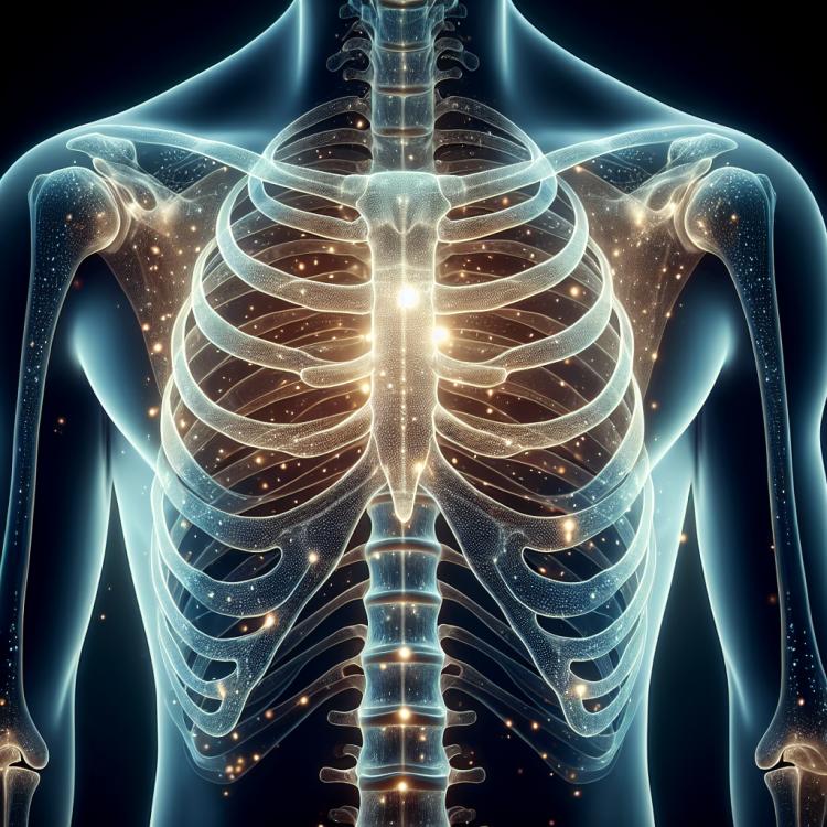
Pectus carinatum: causes of occurrence and treatment methods
- Understanding a keel-shaped chest
- Factors contributing to the development of pectus carinatum
- Characteristics of symptoms of pectus carinatum
- Views of specialists on methods for treating pectus carinatum.
- Methods for diagnosing pectus carinatum
- Methods of treating pectus carinatum
- Preventive measures to prevent pectus carinatum
- Amazing aspects of a keel-shaped chest
- FAQ
Understanding a keel-shaped chest
Pectus excavatum is a congenital defect characterized by a depression or hollow in the chest, usually at the level of the sternum. This condition is associated with an anomaly in the development of the ribs and sternum during the embryonic period, manifested as a concave shape of the chest and can cause cosmetic dissatisfaction and functional problems with breathing or physical exertion.
Understanding pectus excavatum is essential for determining the proper approach to treating this condition. Diagnosis includes clinical examination, chest X-ray, and computed tomography. Treatment can range from conservative methods, such as physical therapy, to surgical correction through plastic surgery or chest reconstruction procedures. It is advisable to consult a specialist to determine the best individualized approach to managing pectus excavatum.
Factors contributing to the development of pectus carinatum
Pectus carinatum may be caused by various factors, including genetic predisposition, pathological anatomy of the ribs or sternum, as well as uneven growth of the ribs and cartilages. Causes may also include inflammatory processes, injuries, or surgical interventions carried out in the chest area. Understanding these factors is crucial for the diagnosis and determination of the optimal treatment plan for pectus deformity.
- Genetic predisposition: Hereditary factors may play a role in the development of pectus carinatum.
- Pathological anatomy of the ribs or sternum: Uneven growth or shape of the ribs and cartilages may also contribute to this condition.
- Inflammatory processes: Infections or inflammations in the chest area may be a contributing factor in the development of the pectus deformity.
- Chest injuries: Damage, wounds, or bruises in the chest area may also influence the formation of pectus deformity.
- Surgical interventions: Surgeries performed in the chest area may sometimes lead to pectus deformity.
Characteristics of symptoms of pectus carinatum
Symptoms of pectus carinatum may include protrusion of the pectus carinatum process of the sternum forward, creating the impression of a “crow’s” chest. Pain and discomfort in the chest area may occur due to asymmetry of the chest or uneven load distribution on the chest wall, which affects the position of the spine. Some patients may also experience respiratory problems due to lung compression from pectus carinatum caused by thoracic compression. The precise characteristics of the symptoms may vary depending on the severity of the deformity and the individual patient’s features.
- Pectus excavatum: this deformity may manifest as a bulge in the chest around the sternum.
- Pain and discomfort: patients may experience pain and discomfort in the chest area due to the unnatural shape and strain on the rib cage.
- Respiratory problems: pectus excavatum may restrict the expansion of the chest during breathing, potentially leading to breathing difficulties.
- Spinal curvature: chest deformity may affect the position of the spine due to uneven load distribution on the back.
- Cosmetic defect: pectus excavatum can cause cosmetic defects, which may affect the psychological state of the patient.
Views of specialists on methods for treating pectus carinatum.
Expert opinion reflects various approaches to the treatment of pectus deformity. Many specialists recommend conservative methods, such as wearing a corset or special devices to correct the position of the chest. The approach is individual and depends on the degree of severity of the deformity in each patient.
However, in complex cases and when there are significant functional or aesthetic problems, surgical intervention may be required. Surgical methods include correcting the shape of the sternum and ribs using implants or rib prostheses. The decision on the treatment method should be made taking into account the individual characteristics of the patient and the potential risks and benefits of performing a specific procedure.

Methods for diagnosing pectus carinatum
Diagnosis of pectus carinatum often includes a visual examination by a doctor, palpation of the chest area to identify anomalies, as well as performing X-rays to visualize abnormalities in the chest structure. For a more detailed assessment of the bones and cartilage of the sternum, additional methods such as computed tomography (CT) or magnetic resonance imaging (MRI) may be used.
Ultrasound examination can also be useful for assessing the internal structures of the chest. In some cases, electrocardiography (ECG) and other methods may be utilized to rule out associated heart or lung problems. Proper diagnosis of pectus deformity is an important step in determining the optimal treatment plan and preventing possible complications.
- Visual inspection and palpation: The doctor conducts an examination and palpation of the sternum to identify any abnormal protrusions or deformities.
- Chest X-ray: Radiographic examination is used to visualize anomalies in the structure of the chest, including bone deformities.
- Computed tomography (CT): A diagnostic imaging method that provides a more detailed image of the structures of the chest and helps determine the nature of the deformity.
- Magnetic resonance imaging (MRI): A diagnostic method that allows for high-quality images of the internal structures of the chest to assess deformities.
- Ultrasound examination: A method that can be used for additional evaluation of the internal structures of the chest and to identify possible anomalies.
Methods of treating pectus carinatum
- Conservative therapy: Includes the use of corsets and special exercises for posture correction and strengthening the chest muscles.
- Surgical correction: In cases of significant deformation and cosmetic defect, surgical intervention may be recommended to correct the shape of the chest.
- Implantation: In some cases, the implantation method is used to normalize the shape of the chest by introducing appropriate prostheses or materials.
- Physiotherapy: Physiotherapeutic procedures can help strengthen muscles and correct the deformity of the chest in cases of pectus carinatum.
- Multimodal approach: A comprehensive approach combining conservative methods, surgery, and physiotherapy is often used for the best treatment results of pectus carinatum.
Preventive measures to prevent pectus carinatum
To prevent pigeon chest, it is recommended to visit a doctor regularly to timely identify any anomalies in the development of the chest. If there is a predisposition to the development of deformities, it is important to discuss preventive measures with a medical professional and follow their recommendations to maintain the health of the chest.
- Maintaining proper posture: Regularly monitoring posture during daily activities and exercises can help prevent the development of pectus carinatum.
- Strengthening back and chest muscles: Various exercises aimed at strengthening the back and chest muscles can contribute to maintaining proper posture and reducing the risk of sternal deformities.
- Avoiding prolonged sitting in incorrect positions: It is especially important for children and adolescents to avoid prolonged sitting in incorrect positions to prevent chest deformities.
- Adhering to a healthy lifestyle: Proper nutrition, physical activity, and overall maintenance of a healthy lifestyle can contribute to strengthening the chest and preventing deformities.
- Regular visits to the doctor: Regular visits to the doctor to monitor the development of the chest and identify anomalies at early stages can help in establishing preventive measures to prevent pectus carinatum.
Amazing aspects of a keel-shaped chest
Although the causes of pectus carinatum deformation can vary, ranging from genetic predisposition to pathological processes, the development and improvement of diagnostic and treatment methods for this condition continue to be an actively studied area in modern medicine. Awareness of pectus carinatum and the ongoing enhancement of its prevention and correction methods help improve the quality of life for patients suffering from this condition.