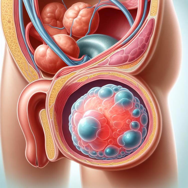
Urachal cyst: causes, diagnosis, and treatment methods
- Understanding Urachal Cyst: Explanation of Symptoms and Causes
- Development of a urachal cyst: pathogenesis and risk factors
- Signs and symptoms of a urachal cyst
- Expert opinion on the treatment of urachal cyst.
- Methods for diagnosing a urachal cyst
- Options for treating a urachal cyst
- Prevention measures for urachal cysts
- Amazing aspects of a urachal cyst
- FAQ
Understanding Urachal Cyst: Explanation of Symptoms and Causes
The Urachal Cyst is an anomaly of formation that arises from the remnants of the urachus – an intrauterine structure connecting the umbilical artery to the bladder. This poorly understood pathology can manifest through various symptoms, including lower abdominal pain, frequent urination, urinary disturbances, and sometimes blood in the urine. The causes of Urachal Cyst formation may be related to genetic factors or pathological development of the urachus during embryonic growth.
For an accurate diagnosis of the Urachal Cyst, comprehensive examination is necessary, including ultrasound of the pelvic organs as well as the urinary tract. Treatment for this condition may include conservative methods, such as observation and medication therapy, or surgical intervention in cases of complications or ineffectiveness of conservative treatment. A clear understanding of the symptoms and causes of Urachal Cyst is important for effective management of this condition.
Development of a urachal cyst: pathogenesis and risk factors
The development of a urachal cyst is associated with an anomaly in embryonic development during the formation of the bladder and urachus. The pathogenesis of the urachal cyst is often related to the formation of cysts from residual tissue cells that usually transform into the urachus. Risk factors include genetic predisposition as well as the presence of other congenital anomalies of the urinary tract, such as a ureterocele or duplication of the urethra. It is important to understand that the exact cause of the urachal cyst’s occurrence is individual for each patient and requires further study.
It should be noted that understanding the pathogenesis and risk factors of the urachal cyst is significant for the diagnosis and treatment of this condition. Further research may help better define the molecular and genetic mechanisms underlying cyst development in patients with this diagnosis, which in turn contributes to a more effective approach to the treatment and prevention of complications of this disease.
- Embryonic development anomaly: the development of a urachal cyst is associated with an anomaly in the formation of the embryo that disrupts the formation of the urachus and bladder.
- Cyst formation from residual cells: the pathogenesis of the urachal cyst is often linked to the formation of cysts from residual cells, which usually develop into the urachus.
- Genetic predisposition: the presence of certain genetic factors may increase the risk of cyst formation in some patients.
- Congenital anomalies of the urinary tract: the presence of other congenital anomalies, such as a kinked ureter or urethral duplication, may also increase the likelihood of urachal cyst formation.
- Research on molecular and genetic mechanisms: further research allows for a better understanding of the molecular and genetic underpinnings of urachal cyst development, improving the diagnostic and treatment options for this condition.
Signs and symptoms of a urachal cyst
Symptoms of a urachal cyst can range from subtle to pronounced, depending on the size and location of the cyst. Patients may present with complaints of tenderness in the abdominal or lumbar area, increased pressure in the bladder, uncontrollable urination, or other signs of urinary dysfunction. It is important to consider that the symptoms of a urachal cyst can mimic other diseases of the urinary system, so differential diagnosis requires extensive clinical analysis.
Detection of a urachal cyst can be performed using various examination methods, including ultrasound, computed tomography, or magnetic resonance imaging. In the case of a confirmed diagnosis, it is important to promptly consult a specialist for appropriate treatment and monitoring of the urachal cyst condition.
- Abdominal or lower back pain: patients with a urachal cyst may experience discomfort or pain in the lower abdomen or lower back.
- Increased pressure in the bladder: a urachal cyst can lead to disruption of normal pressure in the bladder, which may cause discomfort and frequent trips to the restroom.
- Uncontrolled urination: uncontrolled urination or frequent bladder leaks may be a sign of a possible urachal cyst.
- Feeling of pressure or discomfort in the bladder area: some patients may feel pressure or tension in the bladder area due to the presence of a cyst.
- Frequent urinary tract infections: the occurrence of recurrent urinary tract infections may be associated with the presence of a urachal cyst, requiring further examination and treatment.
Expert opinion on the treatment of urachal cyst.
Expert opinions on the treatment of urachal cysts highlight the importance of an individualized approach for each patient. There are several methods for treating urachal cysts, and the choice of the optimal approach depends on factors such as the size of the cyst, the presence of symptoms, and the overall condition of the patient. Many experts believe that observation of small and asymptomatic cysts may be preferable, while larger or symptomatic cysts may require surgical intervention.
Professional opinions also emphasize the importance of regular monitoring of the urachal cyst condition after treatment. Experts recommend conducting follow-up examinations to track treatment effectiveness, changes in cyst size, and to identify potential complications. This approach allows for timely responses to changes and the adaptation of treatment strategies based on the individual characteristics of the patient.

Methods for diagnosing a urachal cyst
To diagnose a urachal cyst, various examination methods are used, including ultrasound, computed tomography (CT), and magnetic resonance imaging (MRI). Ultrasound is a commonly used method that allows visualization of the structure and size of the cyst. Computed tomography (CT) and magnetic resonance imaging (MRI) provide a more detailed view of the location and characteristics of the cyst, which aids in refining the diagnosis and choosing the optimal treatment strategy.
In addition to educational methods, cystoscopy may sometimes be required for a visual examination of the bladder using flexible or rigid endoscopic equipment. The diagnosis of a urachal cyst involves a comprehensive study of the structure and function of the urinary system to establish an accurate diagnosis and determine the optimal treatment plan for each specific case.
- Ultrasound: An important diagnostic method that allows visualization of the urachal cyst and determination of its size and structure.
- Computed Tomography (CT): An advanced method that provides a more detailed image of the cyst and its location in the bladder.
- Magnetic Resonance Imaging (MRI): A diagnostic procedure that helps obtain high-quality images of tissues and organs for accurate identification of the urachal cyst.
- Cystoscopy: An invasive method that can be used for direct visual examination of the bladder to detect and assess the urachal cyst.
- MRI Cystography: A technique that allows assessment of the structure and function of the bladder using magnetic resonance imaging for a detailed study of possible anomalies, including the urachal cyst.
Options for treating a urachal cyst
To achieve optimal results in the treatment of a urachal cyst, it is important to conduct a thorough assessment of the patient, taking into account individual characteristics and medical history. The approach to treatment should be comprehensive, involving a multidisciplinary team of medical specialists, including urologists, surgeons, and radiologists, to determine the most appropriate method of treatment and ensure the best outcome for the patient.
- Dynamic observation: In some cases, small and asymptomatic urachal cysts may not require active treatment and may simply be observed to monitor their dynamics.
- Medication use: To reduce pain, inflammation, or other symptoms related to the cyst, medications such as analgesics or anti-inflammatory drugs may be used.
- Endoscopic removal: An endoscopic procedure may be recommended to remove the cyst through the ureter, which usually helps avoid surgical intervention.
- Open surgical intervention: In the case of large or complicated urachal cysts, surgical intervention may be required to remove the cyst via open surgery.
- Follow-up therapy: After the removal of the urachal cyst, follow-up therapy may be necessary to monitor the patient’s condition and prevent potential recurrence or complications.
Prevention measures for urachal cysts
For the prevention of urachal cysts, it is also important to seek medical assistance in a timely manner when characteristic symptoms or signs of urinary disturbances occur. Regular medical monitoring, especially for individuals with risk factors or a family history, can help identify issues earlier and take appropriate measures to prevent the development of serious conditions related to urachal cysts.
- Healthy Lifestyle: Maintaining a healthy lifestyle, consuming nutrients during pregnancy, and avoiding toxic substances.
- Regular Check-ups and Screenings: Regular visits to the doctor and screenings for pregnant women to identify congenital defects, including urinary system abnormalities.
- Timely Seeking of Medical Help: Seeking medical assistance when characteristic symptoms or signs of urinary disturbances arise.
- Medical Monitoring: Regular medical monitoring of individuals with risk factors or family history to identify problems and prevent the development of a urachus cyst.
- Overall Health Control: Keeping track of overall health status, considering possible hereditary factors and medical history to prevent the development of a urachus cyst.
Amazing aspects of a urachal cyst
Understanding the remarkable aspects of urachal cysts, including risk factors, pathogenesis, and clinical manifestations, is crucial for the timely diagnosis and treatment of this condition. Further research in the field of urinary system medicine may expand our knowledge of urachal cysts and help develop effective treatment strategies for patients facing this rare but significant disease.