Nephroptosis (kidney prolapse): diagnosis, complications, and treatment methods
- Definition and causes of nephroptosis
- Factors contributing to the development of nephroptosis
- Signs and symptoms of nephroptosis
- Expert opinion on the treatment of nephroptosis
- Methods for diagnosing nephroptosis
- Methods of treating nephroptosis
- Measures for the prevention of nephroptosis
- Interesting aspects of nephroptosis
- FAQ
Definition and causes of nephroptosis
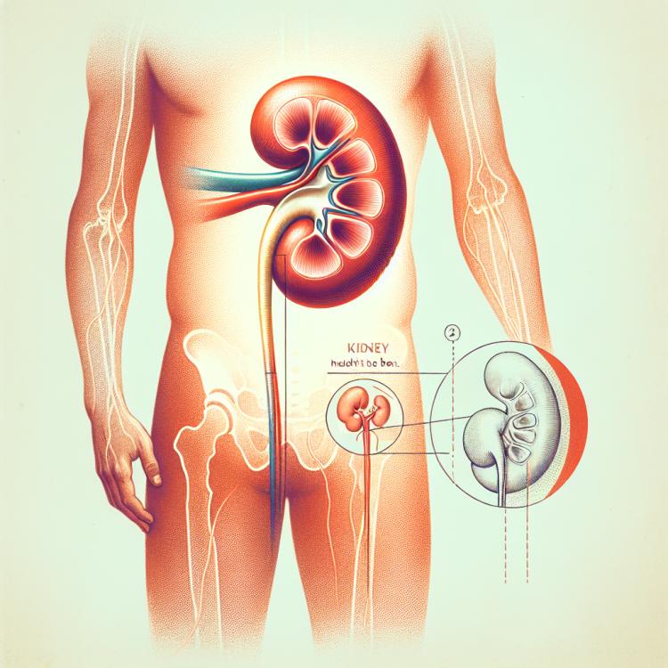
Nephroptosis, also known as kidney prolapse, is a condition in which the kidney is able to descend from its normal position due to insufficient support from the renal tissue stroma and associated ligamentous structures. This can lead to symptoms such as back pain, discomfort, and even impaired kidney function. The origin of nephroptosis is often linked to developmental anomalies, trauma, physical exertion, or significant weight loss, which results in the loss of fatty support in the kidney area and creates conditions for the organ to descend.
Factors contributing to the development of nephroptosis
Nefroptosis, or kidney descent, can be caused by various factors, including ligament abnormalities and atrophy of the kidney parenchyma. Weakness of the abdominal muscles, abdominal injuries, rapid weight loss, and pregnancy can also contribute to this condition. Some patients with nephroptosis have a long surcut, which increases kidney mobility and reduces its fixation in the renal bed, potentially leading to displacement and descent of the kidney.
- Ligament anomalies: Weakness or deformities of the ligaments in the kidney area may contribute to kidney descent.
- Atrophy of the kidney parenchyma: Reduction in the volume of kidney parenchyma may decrease its supportive function.
- Weakness of the abdominal muscles: Insufficient development of the abdominal muscles may not provide adequate support to the kidneys.
- Pregnancy: The increase in the size of the uterus during pregnancy may increase pressure on the kidneys, contributing to their descent.
- Abdominal injuries: Mechanical impact on the abdominal area may cause changes in the position of the kidney.
Signs and symptoms of nephroptosis
Nephroptosis, or kidney dropping, is often accompanied by various symptoms such as pain in the lower back, which may worsen with physical activity or changes in body position. Additionally, patients with nephroptosis may experience a feeling of heaviness or fatigue in the lower back and abdomen.
Other common symptoms of nephroptosis may include urinary disturbances, nonspecific abdominal pain, as well as a general decline in well-being. Patients also often report frequent urination or the opposite phenomenon – slowed urination.
- Back pain: often worsens with physical activity or changes in body position.
- Feeling of heaviness in the lower back and abdomen: patients may experience discomfort and fatigue in these areas.
- Urination disorders: may manifest as increased or decreased frequency of urination.
- Abdominal pain: nonspecific abdominal pain may be one of the signs of nephroptosis.
- General malaise: patients with nephroptosis may experience fatigue, weakness, and other common symptoms of illness.
Expert opinion on the treatment of nephroptosis
Experts in the field of urology and nephrology recommend a comprehensive approach to the treatment of nephroptosis, focusing primarily on addressing the root causes of kidney descent. Frequently used methods may include wearing braces, lifestyle changes, exercises to strengthen muscles, and maintaining the proper position of the kidney. For some patients with more severe cases of nephroptosis, surgical intervention may be required.
Experts also recommend regular monitoring of patients with nephroptosis to track their condition and the effectiveness of treatment. Monitoring symptoms, controlling blood pressure in the renal arteries, and preventive measures are aimed at minimizing the risk of complications and improving the quality of life for patients with nephroptosis.

Methods for diagnosing nephroptosis
To diagnose nephroptosis, doctors may use various methods, including ultrasound of the kidneys, radiological examination with contrast agents, computed tomography, and magnetic resonance imaging. Ultrasound allows for determining the position of the kidney and assessing the degree of its descent, while computed tomography and magnetic resonance imaging provide a more detailed image of the renal fossa.
Clinical symptoms, as well as the results of diagnostic methods, assist doctors in establishing a diagnosis of nephroptosis. Moreover, to confirm the diagnosis, specialists may conduct additional studies using radioisotopes, which allows for a more accurate assessment of the functional state of the kidney and its position.
- Ultrasound examination of the kidneys: A method that allows determining the position of the kidney and assessing the degree of its descent.
- X-ray examination with contrast agents: Helps visualize the kidney and assess its position using contrast media.
- Computed tomography: Provides a more detailed image of the kidney bed area, assisting in diagnosis and assessing the condition of the kidney.
- Magnetic resonance imaging: Offers high-quality images of kidney tissues and surrounding structures for accurate diagnosis of nephroptosis.
- Radioisotope studies: An additional method for assessing the functional state of the kidney and its position to confirm the diagnosis.
Methods of treating nephroptosis
In cases where improvement cannot be achieved through conservative methods, as well as in the presence of serious complications or significant symptoms, surgical intervention may be required. Surgical treatment of nephroptosis may involve fixing the kidney in the correct position with surgical stitches or using special implants to support the kidney.
- Conservative treatment: includes the use of a brace to support the kidney, physical exercises to strengthen the abdominal and lower back muscles, as well as maintaining a normal weight.
- Pharmacological therapy: may be used in rare cases to relieve pain or reduce inflammation in the area of the kidneys.
- Diet and fluid intake: it is recommended to adhere to a healthy diet and sufficient water intake to maintain overall kidney health.
- Surgical treatment: may involve securing the kidney with surgical stitches or implanting special devices to support the kidney in the correct position.
- Rehabilitation and physiotherapy: after treatment, it is important to carry out rehabilitation activities, including physiotherapy exercises to restore muscle and kidney function.
Measures for the prevention of nephroptosis
In addition, one should lead a healthy lifestyle, avoiding excessive strain when lifting weights, regularly undergoing medical check-ups, and following doctors’ recommendations. It is also important to seek medical help promptly when characteristic signs or symptoms appear, in order to rule out possible complications and start treatment in a timely manner.
- Strengthening the abdominal and lower back muscles: Regular physical exercises aimed at strengthening the muscles in the kidney area can help prevent the development of nephroptosis.
- Maintaining a normal weight: Controlling weight and keeping it within normal limits helps reduce the strain on the kidneys and surrounding tissues.
- Avoiding traumatic loads: Preventing traumatic actions on the kidney area, such as heavy physical exertion, helps in the prevention of nephroptosis.
- Adhering to a healthy lifestyle: Avoiding harmful habits, regular sports activities, healthy eating, and optimal hydration will help maintain kidney health.
- Timely consultation with a doctor: If characteristic symptoms appear or if there is suspicion of nephroptosis, it is important to consult a specialist for diagnosis and preventive measures. Early initiation of treatment helps prevent complications.
Interesting aspects of nephroptosis
Moreover, an interesting fact is the variety of diagnostic and treatment methods for nephroptosis that can be applied depending on the individual characteristics of the patient and the degree of progression of the disease. Understanding these aspects helps specialists provide effective treatment and improve the prognosis for patients with this pathology.