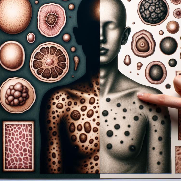
Nevi (moles): causes of occurrence, types, and removal methods
- Information about nevi: their definition and properties
- Etiology of nevi: factors influencing their formation
- Clinical picture of nevi: what you need to know about the symptoms?
- Approaches to the treatment of nevi: expert opinion
- Methods of diagnosing nevi: key aspects and procedures
- Methods for treating nevi: modern approaches and techniques
- Prevention measures for the appearance of nevi: tips and recommendations
- Fascinating aspects about nevi: unexpected facts and features
- FAQ
Information about nevi: their definition and properties
Nevi, also known as moles, are skin formations caused by a buildup of pigment. They can vary in size, shape, and color depending on individual characteristics. Nevi are usually safe, but some of them may undergo changes or be subject to more careful medical monitoring.
Etiology of nevi: factors influencing their formation
Moles, or nevi, are formed due to genetic factors, exposure to ultraviolet rays, and hormonal changes, especially during pregnancy. Genetic mutations can contribute to the formation of nevi, and increased exposure to sunlight can accelerate their appearance on the skin. Additionally, changes in hormone levels in the body can affect the formation of nevi.
- Genetic factors: Hereditary mutations may contribute to the appearance of nevi.
- Ultraviolet radiation: Long-term exposure to ultraviolet rays can accelerate the formation of nevi on the skin.
- Hormonal changes: Fluctuations in hormone levels, especially during periods of pregnancy or adolescence, can provoke the appearance of nevi.
- Mechanical irritation: Friction and pressure on the skin can lead to the formation of nevi in certain areas of the body.
- External injuries: Skin damage, such as burns or wounds, can stimulate the appearance of nevi in these areas.
Clinical picture of nevi: what you need to know about the symptoms?
The clinical picture of nevi can vary depending on their type. Common signs of nevi include spots on the skin of various shapes and sizes, which can be either colored or pigmented. It is important to pay attention to changes in the shape, size, color, or texture of the nevus, as this may indicate a possible malignant transformation. The appearance of itching, bleeding, or changes in the surrounding tissues can also be signs of potentially dangerous changes in the nevus that require further attention and consultation with a specialist.
- Various shapes and sizes: nevi can have a variety of shapes and sizes, ranging from small spots to larger formations on the skin.
- Changes in color and texture: it is important to pay attention to any changes in the color or texture of nevi, as these changes may indicate pathological processes.
- Itching or pain: the appearance of itching or pain in the area of the nevus may require additional attention and examination by a specialist.
- Bleeding: if the nevus starts to bleed without apparent reason, this may also indicate possible problems and requires a doctor’s consultation.
- Changes around the nevus: any changes in adjacent tissues or skin around the nevus, including the formation of new lesions, also require attention and diagnosis.
Approaches to the treatment of nevi: expert opinion
Experts in medicine typically recommend the removal of nevi only in cases where they are suspicious for malignant transformation. However, when choosing a treatment method, various factors are considered, including the type of nevus, its size, location on the patient’s body, and their aesthetic preferences. Commonly used methods for removing nevi include surgical excision, cryodestruction, laser therapy, or electrocoagulation, depending on the individual characteristics of each nevus and the patient’s needs. It is important to have regular examinations with a dermatologist to monitor the condition of existing nevi and identify new ones, in order to take timely measures for their treatment, following the specialists’ recommendations.

Methods of diagnosing nevi: key aspects and procedures
When diagnosing nevi, it is important to conduct a thorough clinical examination, including assessment of the size, shape, color, and texture of the moles. Dermoscopy methods may be used for an accurate diagnosis, allowing for a more detailed study of the structure of nevi and the identification of signs indicating their malignancy. If suspicious changes in the nevi are detected, a biopsy may be required to obtain a tissue sample for subsequent pathological analysis.
- Clinical examination: Includes assessment of the size, shape, color, and texture of the nevi.
- Dermatoscopy: The method allows for viewing the detailed structure of nevi, which helps identify signs of malignant changes.
- Biopsy: The procedure is performed if necessary to obtain a tissue sample of the nevus for pathological analysis.
- Ultrasound examination: The method can be used to assess the structure of nevi and surrounding tissues.
- Computed tomography (CT) or magnetic resonance imaging (MRI): May be used for additional assessment of nevi when malignant processes are suspected.
Methods for treating nevi: modern approaches and techniques
- Laser therapy: One of the modern methods for treating nevi, where a laser is used to reduce the size and pigmentation of moles.
- Surgical removal: Often recommended for nevi suspected of malignancy or for cosmetic reasons.
- Pathological analysis: The removed tissue of the nevus can be subjected to pathological analysis to assess the risk of recurrence and determine the nature of changes in the nevus.
- Liquid nitrogen: Cryotherapy using liquid nitrogen can be applied for deep freezing and removal of nevi.
- Electrocautery: This method can be used to remove nevi by coagulating tissues using electric current.
Prevention measures for the appearance of nevi: tips and recommendations
- Use of sunscreen: Regular application of sunscreen with high SPF helps prevent damage from ultraviolet radiation.
- Limiting time in the sun: Avoid prolonged exposure to direct sunlight during peak hours of solar activity when radiation is most intense.
- Wearing protective clothing: Tight clothing and wide-brimmed hats help protect the skin from direct sunlight.
- Skin examination: Regular skin examinations for changes in existing moles help detect potentially dangerous signs in time.
- Consultation with a dermatologist: Patients with a large number of moles are advised to periodically undergo examinations by a dermatologist for early detection and monitoring of skin condition.
Fascinating aspects about nevi: unexpected facts and features
Another interesting fact is that the appearance of nevuses is related to heredity. People with certain genetic factors may have a greater tendency to develop nevuses. For example, individuals with skin types I and II, who typically have fair skin and light hair, have a higher risk of developing nevuses and, consequently, skin cancer, compared to people with darker skin types.