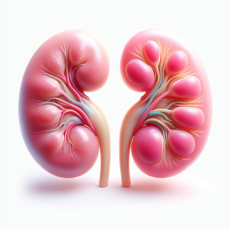
Kidney descent: causes, symptoms, and treatment methods
Dropping of the kidney: main aspects and explanation
The descent of the kidney, or nephroptosis, is a condition in which the kidney shifts below its natural position due to various factors, such as weakness of the ligamentous apparatus or a reduction in the volume of fat tissue in the renal bed. This condition can lead to various symptoms, including lower back pain, frequent urination, and even urinary dysfunction.
Understanding the main aspects of kidney descent is important for the diagnosis and treatment of this condition. In this regard, studying the mechanisms that lead to the development of nephroptosis, as well as methods of diagnosis and treatment, contributes to a more effective approach to managing this condition in patients.
Etiology of kidney ptosis
Prolapse of the kidney or nephroptosis is a condition in which the kidney shifts from its normal position in the lumbar region of the abdominal cavity due to a reduction in the ligaments that hold the kidney in its upper location. Contributing factors include abnormalities in the structure of the kidney’s supportive tissues, atrophy of the fatty layer that usually allows the kidney to remain in place, as well as injuries or strains of the childbirth ligaments.
The main causes of kidney prolapse can be atrophy of the liver capsule, disruption of the usual positioning of blood circulation, chronic peritonitis, systemic disease syndrome, as well as severe drooping of the large and small intestines.
- Defects in the structure of support tissues: Congenital anomalies or defects in the ligamentous or capsular structures of the kidney can make it more prone to displacement.
- Atrophy of the fatty layer: A decrease in the volume of fatty tissue surrounding the kidney may reduce its supportive properties, contributing to its descent.
- Injuries and loads: Injuries or strains to ligaments, for example, as a result of heavy lifting, can damage the structures that hold the kidney in its normal position.
- Hypotrophy of the liver capsule: A reduction in the volume or thickness of the liver capsule may be accompanied by a decrease in the support of the kidney in its upper location.
- Systemic disease syndrome: Some systemic diseases can lead to changes in connective tissue, which contributes to the descent of the kidney.
The clinical picture of kidney prolapse
The clinical picture of kidney displacement can manifest through various symptoms, including back or abdominal pain, frequent urination, blood in the urine, tenderness in the kidney area upon palpation, as well as changes in the size and position of the kidney during ultrasound and other examinations. Patients may also experience malaise, fatigue, and general discomfort due to functional disorders associated with kidney displacement.
It is important to note that symptoms may vary depending on the degree of kidney displacement, the presence of concomitant diseases, and individual characteristics of the body. Diagnosing kidney displacement includes conducting a physical examination, laboratory and instrumental studies that will allow establishing a diagnosis and determining the best treatment methods for this pathological condition.
- Back or abdominal pain: patients with kidney ptosis often complain of discomfort, pain, or pressure sensations in the back or abdomen.
- Frequent urination: changes in the position of the kidney may exert pressure on the bladder, leading to frequent trips to the restroom.
- Blood in urine: the presence of blood in the urine, or hematuria, can be one of the signs of impaired kidney function due to ptosis.
- Tenderness on palpation: during the examination of the kidney area, patients may feel tenderness or discomfort at the site of the kidney ptosis.
- General malaise: patients with kidney ptosis may experience general malaise, fatigue, and weakness due to impaired kidney function and discomfort in the back.
Expert opinions on the treatment of kidney prolapse
Experts in the field of urology and nephrology pay special attention to the individual approach to the treatment of kidney prolapse, depending on the degree of prolapse, clinical manifestations, possible complications, and the general condition of the patient. Based on this, the optimal treatment method is chosen considering all the aforementioned factors to ensure the best results and minimize possible complications.
Expert opinion emphasizes that conservative treatment methods, such as physical exercises to strengthen the abdominal and lower back muscles, as well as wearing a support belt, can be effective in the early stages of kidney prolapse. However, in more advanced cases, surgical intervention, such as nephropexy, may be required to restore the normal position of the kidney and prevent further complications.

Diagnosis of kidney ptosis
Various methods are used for the diagnosis of kidney descent, including a physical examination of the patient to assess symptoms and complaints, performing an ultrasound of the kidneys to determine the position and condition of the kidney, as well as MRI diagnostics, which provide a more detailed image of the abdominal organs and possible complications. Additionally, laboratory tests such as a complete blood count and urinalysis may be required to identify possible changes associated with kidney descent.
It is important to emphasize that accurate diagnosis of kidney descent is a key step in determining the optimal treatment plan. Timely detection of the pathology and assessment of the degree of descent enable medical professionals to develop a personalized approach for each patient and choose the most effective methods for correcting this condition.
- Physical examination: The doctor conducts an examination of the patient and palpation of the kidney area to identify tenderness or changes in the position of the kidney.
- Kidney ultrasound: Ultrasound allows for determining the position, size, and structure of the kidneys, as well as identifying the presence of nephroptosis.
- MRI diagnostics: Magnetic resonance imaging provides a more detailed image of kidney tissues, which helps clarify the diagnosis and identify possible complications.
- Laboratory tests: A general blood and urine test allows for the identification of changes associated with nephroptosis, such as the presence of protein or blood in the urine.
- Computed tomography (CT): Computed tomography provides more accurate information about the structure and position of the kidneys, aiding in the diagnosis of nephroptosis.
Treatment of kidney descent
Surgical treatment methods, including resection of the excess length of the renal pedicle or reinforcement of the kidney using special materials, may be recommended in cases where conservative methods prove insufficiently effective. The decision on the choice of treatment method should be made by the physician based on the individual characteristics of the patient and the disease, taking into account the minimization of risks and the best clinical outcomes.
- Therapeutic exercise: physical exercises aimed at strengthening the pelvic and abdominal muscles can be effective for improving kidney support.
- Wearing elastic bands: special supportive devices can help keep the lowered kidney in place and reduce discomfort.
- Lifestyle changes: adjusting diet, avoiding excessive physical strain, and maintaining a healthy lifestyle can contribute to the improvement of the condition of a dropped kidney.
- Pharmacological treatment: in some cases, medications may be prescribed to alleviate symptoms such as pain or dysuria associated with a dropped kidney.
- Surgical intervention: in severe cases of kidney descent, when conservative methods do not yield the expected results, surgery may be required to restore the normal position and function of the kidney.
Prevention of kidney prolapse
In addition, it is important to have regular medical check-ups, especially when there are risk factors for developing kidney descent, such as pregnancy or excess weight. If symptoms or changes predisposing to this condition are detected, the doctor may recommend additional prevention measures or monitor kidney condition to prevent descent.
- Regular physical exercise: Strengthening the pelvic floor and abdominal muscles helps maintain the proper position of the kidneys and prevents their descent.
- Maintaining the correct posture during urination: Avoiding excess strain while urinating will help prevent additional pressure on the kidneys.
- Maintaining a healthy weight: Weight management helps reduce the burden on the kidneys and prevents the risk of their descent.
- Mindful weight lifting: When lifting weights, it is important to maintain the proper body position and avoid excessive strain to not overload the kidneys.
- Regular medical check-ups: Regular visits to the doctor can help identify predisposing factors for kidney descent and take measures to prevent it.
Interesting facts about kidney dropping
Another interesting aspect is that the risk of kidney descent may increase with the presence of obesity, excessive strain, or excessive physical stress on the abdominal cavity. Understanding these factors and their impact on the development of kidney descent allows doctors and patients to take measures to prevent this condition and provide more effective treatment.