Osteoma of the frontal sinus: symptoms, causes, and treatment
- Understanding frontal sinus osteoma: key aspects
- Etiology of frontal sinus osteoma
- Clinical picture of frontal sinus osteoma
- Expert opinion on the treatment of frontal sinus osteoma
- Methods for diagnosing frontal sinus osteoma
- Approaches to the treatment of frontal sinus osteoma
- Prevention of frontal sinus osteoma
- Amazing facts about frontal sinus osteoma
- FAQ
Understanding frontal sinus osteoma: key aspects
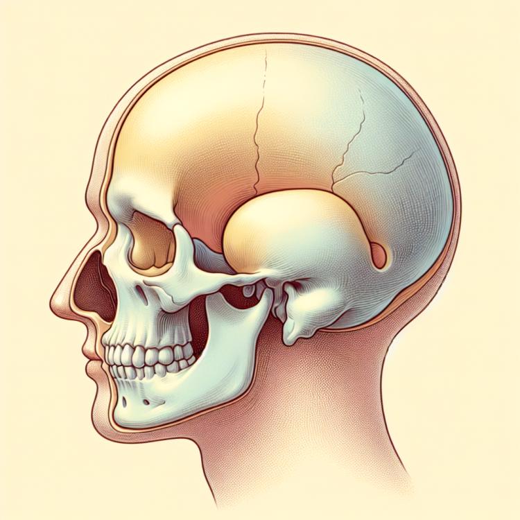
The frontal sinus osteoma is a benign tumor that usually does not cause symptoms. This tumor consists of bone tissue and may be found accidentally during an examination of the frontal sinus for other reasons. In most cases, the frontal sinus osteoma does not require treatment and is a monitored condition; however, in rare cases, surgery may be needed if symptoms arise, such as headaches, nasal congestion, or other problems related to the tumor.
Etiology of frontal sinus osteoma
The etiology of frontal sinus osteoma includes several factors. One of the main ones is genetic predisposition, which may play a role in the development of this tumor formation. Additionally, chronic inflammatory processes in the area of the frontal sinus can contribute to the formation of osteoma.
Other possible causes include facial area injuries, congenital anomalies of skull bone development, as well as radiation exposure. It is important to consider all these factors when discussing the etiology of frontal sinus osteoma and to approach diagnosis and treatment with regard to potential causes to ensure the best outcomes for the patient.
- Genetic predisposition: a family history of frontal sinus osteoma may increase the risk of developing this disease.
- Chronic inflammatory processes: prolonged inflammation in the area of the frontal sinus may contribute to the formation of an osteoma.
- Facial injuries: injuries and damage, especially in the area of the facial bones, may be associated with the development of an osteoma.
- Congenital anomalies of skull bone development: anomalies in the development of skull bones may predispose to the occurrence of osteoma.
- Radiation exposure: prolonged or repeated exposure to radiation in the head and neck area may be associated with the development of frontal sinus osteoma.
Clinical picture of frontal sinus osteoma
The clinical picture of an osteoma of the frontal sinus may manifest with various symptoms, including chronic nasal congestion, a sensation of pressure in the facial area, as well as abundant mucous or mucopurulent nasal discharge. Patients may also experience pain or discomfort in the forehead area, above the eyes, or around the nose.
Other symptoms may include a deterioration of smell, a decreased ability to distinguish odors, and a possible development of chronic sinusitis. It is important to pay attention to these signs when suspecting an osteoma of the frontal sinus and to conduct appropriate diagnostics to establish an accurate diagnosis and develop a treatment plan.
- Chronic nasal congestion: patients may experience a feeling of constant nasal congestion, which can complicate normal breathing through the nose.
- Pressure in the facial area: the pressure that arises in the facial area, especially in the forehead or above the eyes, can be one of the characteristic symptoms of a frontal sinus osteoma.
- Mucous or mucopurulent nasal discharge: the presence of abundant mucous or purulent discharge from the nose may indicate the presence of an osteoma and an accompanying inflammatory process.
- Pain in the facial area: pain or discomfort in the facial area, especially in the forehead, above the eyes, or around the nose, can be one of the symptoms of a frontal sinus osteoma.
- Deterioration of smell: patients may experience changes in smell, up to a complete loss of the ability to recognize odors, which may be related to a frontal sinus osteoma and its impact on the olfactory nerves.
Expert opinion on the treatment of frontal sinus osteoma
Expert opinion on the treatment of frontal sinus osteoma is based on a comprehensive approach to managing this condition. Experts recommend a personalized approach to planning the treatment strategy, taking into account the size and location of the tumor, the presence of symptoms, and the overall health of the patient. Surgical removal of the osteoma may be recommended in cases where the tumor causes significant symptoms or poses a threat to surrounding tissues.
Furthermore, experts emphasize the importance of regular monitoring and follow-up after treatment to prevent potential recurrences or complications. They also highlight the significance of adhering to all specialists’ recommendations and regular medical consultations to achieve the best outcomes and prevent possible complications.

Methods for diagnosing frontal sinus osteoma
Various examination methods are used for the diagnosis of frontal sinus osteoma. Imaging techniques, such as X-rays, computed tomography (CT), and magnetic resonance imaging (MRI), provide detailed images of bone tissue anomalies in the area of the frontal sinus. These methods help to determine the size and nature of the osteoma, as well as its relation to the surrounding tissues.
Additionally, conducting rhinoscopic investigations, such as rhino-fibroscopy and endoscopy of the nasopharynx, may be useful for a more detailed assessment of the mucous membrane and possible involvement of blood vessels or nerves. Combining various diagnostic methods allows for an accurate diagnosis of frontal sinus osteoma and the determination of an optimal treatment plan for the patient.
-
– X-ray: This method involves the use of X-rays to create an image of the bony structure of the frontal sinus, allowing for the determination of the presence of a tumor or bone anomalies.
– Computed Tomography (CT): CT scanning provides more detailed three-dimensional images of the frontal sinus area, which helps determine the size, shape, and location of the osteoma.
– Magnetic Resonance Imaging (MRI): MRI allows for high-quality images of tissues and structures in the head and neck area, aiding in the diagnosis of osteoma of the frontal sinus.
– Rhinofibroscopy: This method allows the physician to examine the nasal cavity and frontal sinus using a flexible endoscope, which can help identify possible changes caused by the osteoma.
– Nasopharyngoscopy: Conducting an endoscopy can be a valuable method for further examining the mucosa and assessing the possible involvement of surrounding tissues in the development of the osteoma of the frontal sinus.
Approaches to the treatment of frontal sinus osteoma
In the case of small and asymptomatic osteomas, the patient may be monitored by specialists with regular examinations to monitor the state of the tumor. In more serious cases, when the tumor causes discomfort or compresses surrounding tissues, surgical intervention may be necessary to remove the osteoma and restore normal anatomical structure.
- Observation and dynamic observation: For patients with small osteomas or without obvious symptoms, a decision may be made to conduct regular observation and monitor the condition of the tumor.
- Medication therapy: In some cases, medications may be prescribed to reduce symptoms such as pain or inflammation related to the frontal sinus osteoma.
- Surgical removal of the tumor: In the presence of large or symptomatic osteomas, surgery may be required for complete or partial removal of the tumor.
- Reconstructive surgery: In cases where the osteoma leads to deformation or damage to surrounding tissues, reconstructive surgical interventions may be necessary to restore anatomical integrity.
- Combination of treatment methods: Sometimes specialists may apply a combined approach, including observation, medication therapy, and surgical intervention, depending on the characteristics of each specific case.
Prevention of frontal sinus osteoma
Preventing injuries and external influences on the facial area, as well as maintaining a healthy lifestyle that includes a balanced diet, adequate physical activity, and avoiding toxic substances, can also help reduce the risk of developing frontal sinus osteoma.
- Regular consultations with a doctor: it is important to see a specialist at the first signs of diseases in the facial area.
- Avoiding traumatic impacts: preventing injuries and bruises in the facial area helps to protect anatomical structures.
- Keeping a healthy lifestyle: leading an active lifestyle, proper nutrition, and giving up bad habits help maintain overall health.
- Preventing exposure to harmful factors: refraining from smoking, protecting against radiation and chemicals contribute to maintaining the health of bone tissue.
- Seeking medical help when necessary: timely diagnosis of potential issues and immediate treatment help prevent the development of frontal sinus osteoma.
Amazing facts about frontal sinus osteoma
Interestingly, the osteoma of the frontal sinus is usually a benign tumor and has a slow progression. However, in rare cases, the osteoma can lead to various symptoms, such as nasal congestion, facial pain, and other manifestations that require the intervention of specialists for diagnosis and treatment.