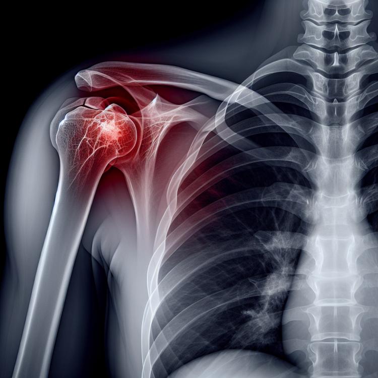
Fracture of the radial head: signs, causes, and treatment methods.
- Understanding a fracture of the radial head
- Etiology of radial head fracture
- The clinical picture of a fracture of the radial head
- Expert opinion on the treatment of a radial head fracture
- Methods for diagnosing a fracture of the radial head
- Methods of treating a fracture of the radial head
- Measures for the prevention of radial head fracture
- Amazing aspects of radial head fracture
- FAQ
Understanding a fracture of the radial head
A fracture of the radial head is an injury in which the bony prominence at the top of the radius tears or breaks. This type of fracture often occurs as a result of trauma, such as falling on an outstretched arm. The radial head plays an important role in the stability and functioning of the wrist, so a fracture in this area can significantly affect the overall functionality of the hand. Treatment of a radial head fracture may include conservative methods, such as splint immobilization, or surgical intervention, especially in cases of severe injury or displacement.
Etiology of radial head fracture
A fracture of the radial head can occur as a result of trauma or injury, most often due to a fall onto an outstretched arm or a direct blow to the forearm. This type of fracture is commonly seen in athletes and patients engaged in physical activity. Additionally, the disruption of bone structure can be caused by osteoporosis or other musculoskeletal disorders, making the bone more susceptible to fractures.
- Injury: a fracture of the radial head can occur as a result of falling on an extended arm or a direct blow to the forearm.
- Sports injuries: athletes and people engaged in physical activity are at risk of fracturing the radial head.
- Osteoporosis: a condition in which bones become brittle and more susceptible to fractures may contribute to a fracture of the radial head.
- Musculoskeletal disorders: a number of diseases, such as osteomalacia, can increase the risk of a radial head fracture.
- Repetitive injuries: frequent repetitive injuries in the forearm area can contribute to the development of a radial head fracture.
The clinical picture of a fracture of the radial head
The clinical picture of a radial head fracture usually includes severe pain in the forearm, swelling, bruising, and limited mobility in the elbow joint. Patients may also experience tingling or numbness in the forearm and fingers due to nerve endings being compressed during the fracture.
To confirm the diagnosis of a radial head fracture, X-ray imaging is necessary. Symptoms may also include cracking sounds when attempting to flex and extend the elbow. Treatment typically involves wearing an elbow brace, physical therapy, and in some cases, surgical intervention.
- Severe pain: patients experience intense pain in the forearm area associated with a fracture of the radial head.
- Swelling and bruising: tissue swelling and the appearance of bruises at the injury site are observed, related to the inflammatory response to the damage.
- Limited mobility: patients may have difficulty moving the elbow joint due to pain and bone deformation from the fracture.
- Tingling and numbness: tingling or numbness may occur in the forearm and fingers due to nerve compression.
- Cracking during movement: patients may report cracking or creaking in the elbow joint when attempting to bend or straighten the arm due to the disruption of bone integrity.
Expert opinion on the treatment of a radial head fracture
The expert opinion on the treatment of a radial head fracture is based on the need for an individualized approach for each patient, taking into account the characteristics of the injury, the presence of complications, and other factors. Specialists in orthopedics and surgery adhere to the principle of maximizing function preservation and restoring bone structure when treating such injuries.
Many experts emphasize the importance of early diagnosis and timely treatment of a radial head fracture, which contributes to accelerating the healing process and reducing the risk of complications. Experts may also suggest innovative treatment methods, such as surgical correction in complex fractures or the use of physiotherapy for joint function recovery.

Methods for diagnosing a fracture of the radial head
To diagnose a fracture of the radial head, the doctor typically conducts a physical examination of the patient, assessing pain, swelling, and mobility in the elbow area. To confirm the diagnosis, the doctor may recommend X-rays, which allow visualization of the bone damage and determine its exact position.
In the presence of complex or ambiguous cases of radial head fractures, the doctor may prescribe additional examinations, such as computed tomography (CT) or magnetic resonance imaging (MRI), to obtain more detailed information about the damage and its impact on the joint structures.
- Physical Examination: the doctor conducts an examination, evaluates symptoms, and checks the mobility in the elbow area to identify signs of a fracture of the radial head.
- X-ray: the primary method for diagnosing bone fractures, which allows viewing images of bone structure and determining the presence and nature of damage.
- Computed Tomography (CT): a more detailed imaging method that may be prescribed for a more precise determination of the nature of the fracture.
- Magnetic Resonance Imaging (MRI): allows obtaining images of soft tissues and joints, which can be useful for additional diagnostics in complex fracture cases.
- Ultrasound Examination: sometimes used to assess soft tissues in the area of the injury, especially in cases where other methods are insufficient for diagnosis.
Methods of treating a fracture of the radial head
In some cases, surgical intervention may be required to restore stability and proper bone anatomy. The surgeon may use internal fixation methods, such as osteosynthesis with metal plates and screws, to ensure proper and rapid rehabilitation of the patient.
- Application of an elbow brace: to stabilize and reduce load on the injured area.
- Physical therapy: prescribed to restore joint mobility and strengthen surrounding muscles and ligaments.
- Surgical intervention: sometimes required to restore stability and correct bone anatomy.
- Osteosynthesis with metal plates and screws: used to ensure proper rehabilitation of the patient.
- Rehabilitation activities: include controlled exercises to restore joint functionality and strengthen muscles.
Measures for the prevention of radial head fracture
It is also important to follow safety precautions when engaging in sports or other types of physical activity to avoid falls or injuries that could lead to a radial head fracture. Regular consultations with a doctor to evaluate bone health, especially if there are risk factors such as osteoporosis, also contribute to fracture prevention.
- Physical activity: Regular exercises to strengthen muscles and bones help increase the overall strength of bone tissue, reducing the likelihood of fractures.
- Avoiding risky situations: Precautionary measures during sports or other forms of physical activity help prevent falls and injuries that could lead to a fracture of the radial head.
- Calcium-rich diet: Consuming foods that contain a sufficient amount of calcium contributes to strengthening bones and may reduce the risk of fractures.
- Preventive consultations with a doctor: Regular check-ups to assess the overall condition of bones, especially when there are risk factors such as osteoporosis, help timely diagnose and prevent potential problems.
- Study and adherence to individual recommendations: For individuals at increased risk of radial head fractures or other bone problems, it is important to follow the individual recommendations of the doctor and adhere to preventive advice.
Amazing aspects of radial head fracture
Moreover, studies show that athletes and people engaged in an active lifestyle have an increased risk of suffering radial head fractures due to excessive loads on the bone tissue. Therefore, it is important to consider preventive measures and conduct regular examinations for the timely detection and treatment of such injuries.