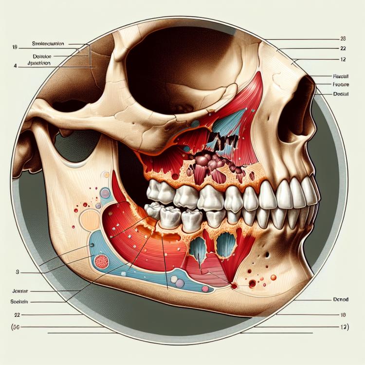
Zygomatic bone fracture: diagnosis, treatment, and rehabilitation
- Definition of zygomatic bone fracture
- Predisposing factors for zygomatic bone fracture
- Signs of zygomatic bone fracture
- The specialists’ vision on the therapy of zygomatic bone fracture
- Methods for diagnosing a zygomatic bone fracture
- Therapy for zygomatic bone fracture
- Measures to prevent zygomatic bone fracture
- Unusual aspects of zygomatic bone fracture
- FAQ
Definition of zygomatic bone fracture
A zygomatic bone fracture is a damage to the bony structure characterized by a rupture of the bone’s integrity and is accompanied by a displacement of its fragments. Zygomatic bone fractures can occur due to trauma, a blow, or an accident, leading to significant injury to the facial area. Diagnosis of a zygomatic bone fracture is based on clinical signs, X-ray examination, as well as computed tomography for accurate determination of the characteristics of the injury.
Predisposing factors for zygomatic bone fracture
Various factors can predispose to a zygomatic bone fracture. Traumatic situations such as car accidents, falls from height, and blows to the face during sports are considered one of the main causes of this type of injury. Additionally, certain medical conditions like osteoporosis can reduce bone density and increase the risk of fractures, including zygomatic bone fractures. It is important to consider these factors when determining possible causes and preventing such injuries.
- Traumatic situations: car accidents, falls from heights, and blows to the face during sports can be predisposing factors for zygomatic bone fractures.
- Medical conditions: low bone density due to osteoporosis or other bone diseases can increase the risk of fractures in this part of the skull.
- Genetic predisposition: the presence of congenital or inherited bone abnormalities can increase the likelihood of zygomatic bone fractures.
- External impacts: shock waves, direct blows to the zygoma, as well as the effects of sharp or blunt objects can contribute to bone fractures.
- Injuries from sports activities: accidents during sports or contact sports can lead to zygomatic bone fractures as a result of facial trauma.
Signs of zygomatic bone fracture
The symptoms of a facial bone fracture can vary depending on the nature of the injury. Typically, a facial bone fracture presents with tenderness and swelling in the area of the injury, disruption of the skin’s integrity, bruising, as well as puffiness and discoloration. Patients may also experience changes in the shape of the face or unevenness of the cheekbones. When these signs appear, it is important to consult a physician for further examinations and appropriate treatment.
- Pain and swelling: usually, with a fracture of the cheekbone, tenderness is observed upon touching the damaged area, as well as swelling.
- Disruption of the integrity of the skin: disruption of the skin integrity in the area of injury may be a sign of a cheekbone fracture.
- Hemorrhages: hemorrhages in the area of injury may occur, which can worsen swelling and skin discoloration.
- Swelling and bruises: a cheekbone fracture is often accompanied by swelling and the appearance of bruises in the area of damage.
- Change in facial shape: sometimes a fracture of the cheekbone can lead to a change in facial shape or visible asymmetry.
The specialists’ vision on the therapy of zygomatic bone fracture
Experts in the field of orthopedics and traumatology emphasize the importance of a comprehensive approach to the treatment of zygomatic bone fractures. One of the main methods of therapy is surgical intervention, which allows for the restoration of normal bone anatomy and reduces the risk of complications. Experts also recommend rehabilitation after surgery to restore functionality in the facial area and improve the patient’s quality of life.
The specialists’ approach to treating zygomatic bone fractures is based on current scientific research, clinical experience, and the individual characteristics of each patient. They emphasize the necessity of a personalized approach and the assessment of potential risks and benefits from various treatment methods. Experts strive to provide patients with the best quality of care and treatment outcomes aimed at restoring facial skeleton functionality and preventing complications.

Methods for diagnosing a zygomatic bone fracture
The diagnosis of a zygomatic bone fracture typically includes a clinical examination of the patient with an assessment of symptoms and preceding traumatic situations. X-ray examination is the primary method of investigation to confirm the diagnosis, allowing visualization of the fracture’s presence, type, and characteristics, such as fragment displacement. In some cases, additional imaging methods, such as computed tomography or magnetic resonance imaging, may be required for a more detailed assessment of the damage and treatment planning.
- Clinical examination: The doctor conducts an examination, determines the presence of tenderness, swelling, bruising, and other signs of a fracture.
- X-ray: One of the primary diagnostic methods that allows visualization of bone injuries and determines their characteristics.
- Computed Tomography (CT): An additional study that provides more detailed images of bone structures for accurate diagnosis.
- Magnetic Resonance Imaging (MRI): Used for more detailed visualization of soft tissues and adjacent structures, critical in facial bone fractures.
- Ultrasound of facial bones: This method may be applicable in diagnosing fractures of the zygomatic bone, especially in children or when assessing soft tissues is necessary.
Therapy for zygomatic bone fracture
- Depending on the nature and severity of the fracture, both conservative treatment (simple fractures without displacement) and surgical treatment (displaced and complex fractures) may be applied.
- Conservative therapy includes wearing special devices and orthoses to stabilize the bone in place and prevent additional stress during healing.
- Surgical treatment may involve reduction – restoring the anatomical position of bone fragments, as well as fixation using plates, screws, or other implants.
- After treatment, it is important to provide the patient with adequate rehabilitative care and physiotherapy to restore the functionality of the damaged bone and surrounding tissues.
- Regular monitoring of the treatment process and X-ray examinations will help assess the effectiveness of therapy and take necessary measures for the successful healing of the fracture.
Measures to prevent zygomatic bone fracture
- Compliance with safety measures: Avoid dangerous situations that may lead to falls or blows to the face and head.
- Wear protective gear: When engaging in sports, especially contact sports, use helmets or other head protection.
- Maintain bone density: A balanced diet rich in calcium and vitamin D helps strengthen bones and may reduce the risk of fractures.
- Avoid dangerous situations on the roads: Be attentive while driving and follow road safety rules to prevent accidents that may lead to head injuries.
- Preventive measures when working at heights: When working at heights, use special protective equipment and follow safety instructions to prevent falls and possible head and face injuries.