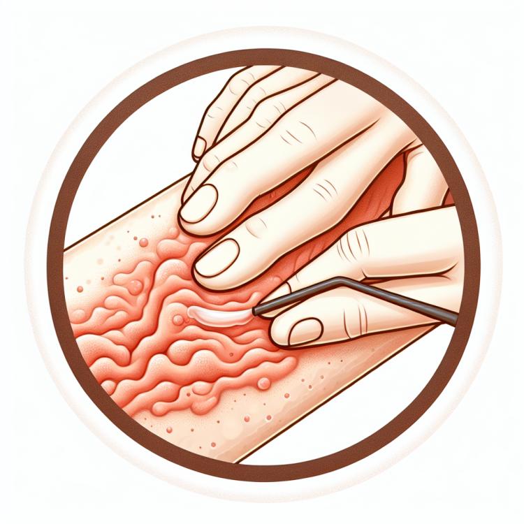
Seroma: possible causes and effective treatment
Understanding Seroma: Key Aspects and Description
Seroma is a formation in the body’s cavity filled with serous fluid. The main aspects of understanding seroma are its possible causes and the mechanism of formation, as well as the diagnosis and treatment of this condition. The description of seroma includes various types of this pathological state, possible symptoms and consequences, as well as methods of diagnosis and therapy aimed at the successful treatment of this disease.
Etiology of Seroma
Seroma is an abnormal formation that occurs as a result of the accumulation of lymphatic fluid or serous fluid in the tissues of the body. The causes of seroma development can include trauma, surgical interventions, infections, diseases of the lymphatic system, or follicular cysts. Other factors contributing to the appearance of seroma may include increased pressure in the tissues or changes in lymphatic flow caused by various pathological processes in the body.
- Injuries: physical damage to tissues can lead to disruption of lymphatic flow and the formation of a seroma.
- Surgical interventions: postoperative complications can be one of the causes of seroma development.
- Infections: inflammatory processes can lead to the accumulation of lymph or serous fluid, forming a seroma.
- Lymphatic system diseases: dysfunctions in the lymphatic system can be a risk factor for seroma.
- Follicular cysts: various cystic formations can lead to the formation of a seroma in the body.
Clinical picture of Seroma
The clinical manifestations of a seroma can vary depending on its location and size. The main symptoms of a seroma include swelling, the formation of a bulge or tumor in the area of fluid accumulation, a sense of tightness and pressure in the affected area, sometimes accompanied by tenderness. Patients may also experience discomfort or a feeling of heaviness around the formation, which can lead to a deterioration in the quality of life. In the presence of a seroma in the lungs, the development of cough, shortness of breath, and other respiratory symptoms may occur, necessitating timely diagnosis and treatment.
- Swelling in the area of fluid accumulation, usually accompanied by an increase in the volume of the affected tissue.
- Formation of a bulge or tumor that may be visible or felt upon palpation.
- Sensation of tension and pressure at the site of seroma formation.
- Possible impairment of the function of nearby organs and tissues caused by pressure from the accumulated fluid.
- In some cases, the appearance of tenderness or discomfort at the site of seroma localization.
Expert opinion on the treatment methods for seroma
Expert opinion on the methods for treating seromas emphasizes the importance of an individualized approach for each patient. Determining the best treatment method depends on the size and location of the formation, overall health status, and possible complications. Experts may recommend conservative methods, such as fluid drainage, the use of compression bandaging, or medication therapy, or surgical intervention in cases where other methods prove ineffective or insufficiently safe.

Methods of diagnosing Seroma
Diagnosis of seroma includes the use of various examination methods such as physical examination, ultrasound examination, computed tomography, magnetic resonance imaging, and puncture biopsy. Physical examination allows the doctor to assess the size, shape, and consistency of the formation, as well as evaluate accompanying symptoms. Ultrasound examination provides detailed images of internal tissues and assesses the presence of fluid in the formation.
Computed tomography and magnetic resonance imaging are used to obtain a more detailed picture of the affected area, determining the structure, distribution, and connections of the formation. Puncture biopsy can be performed to obtain a tissue sample for laboratory analysis and clarification of the diagnosis. The comprehensive application of various diagnostic methods allows for an accurate determination of the nature of the seroma, which is important for choosing the optimal treatment method.
- Physical examination: The doctor performs a visual inspection and palpation of the formation to assess its characteristics, size, shape, and symptoms.
- Ultrasound examination: This method provides a detailed image of internal tissues, evaluating the volume and structure of the formation, as well as the presence of fluid within it.
- Computed tomography (CT): CT provides more detailed imaging of the affected area, detecting structural details of the formation and its distribution.
- Magnetic resonance imaging (MRI): MRI accurately displays soft tissues, allowing assessment of the composition of the formation and its relationships with surrounding structures.
- Fine-needle aspiration biopsy: A fine-needle aspiration of the formation may be performed to obtain a tissue sample for laboratory analysis and to clarify the diagnosis.
Methods of treating Seroma
For larger seromas that cause symptoms or discomfort, drainage, sclerotherapy, or surgical intervention methods may be applied. Drainage allows for the removal of accumulated fluid using a needle or catheter. Sclerotherapy can be used to close the cavity of the seroma and prevent its reformation. In some cases, surgical removal of the seroma is required to prevent complications or recurrences.
- Conservative observation and waiting: In the case of small and asymptomatic seromas, a wait-and-see approach can be used, as some of them may resolve on their own.
- Medication therapy: The use of medications, such as diuretics, can help reduce fluid accumulation in the tissues.
- Drainage: A drainage procedure can be performed to remove accumulated fluid using a needle or catheter under the guidance of ultrasound or other medical education methods.
- Sclerotherapy: The sclerotherapy procedure allows for the closure of the seroma cavity by injecting a special substance that promotes tissue adhesion and prevents fluid re-accumulation.
- Surgical removal: If necessary and in the presence of large seromas causing symptoms or complications, surgical removal of the formation may be required to prevent recurrences and further complications.
Prevention measures for seroma
It is also important to closely monitor the rehabilitation process after surgeries, paying attention to the manifestation of signs of inflammation or other adverse symptoms. Regular consultations with a doctor and adherence to individual medical recommendations will help timely identify and prevent possible complications, including the development of seroma.
- Proper wound care: Regular and proper care of wounds after injuries or surgical interventions reduces the risk of seroma development.
- Following medical recommendations: It is important to follow doctors’ instructions regarding drain care and rehabilitation guidelines after surgeries to prevent the occurrence of seroma.
- Adequate drainage provision: Ensuring proper and effective drainage after surgeries helps prevent fluid accumulation and seroma development.
- Avoiding injury to the area: Patients are advised to avoid over-bandaging, chafing, or injuring the area where a seroma may form to prevent its development.
- Careful monitoring of symptoms: It is important for patients to monitor their overall condition after surgeries and promptly seek medical attention if signs of inflammation or other adverse symptoms arise.