Synostosis: diagnosis, treatment, and consequences
- Understanding Synostosis: Basics and Essence
- Pathophysiology of synostosis
- Clinical picture of synostosis
- Expert opinion: approaches to the treatment of synostosis
- Methods of diagnosing synostosis
- Approaches to the treatment of synostosis
- Prevention measures for synostosis
- Amazing aspects of synostosis
- FAQ
Understanding Synostosis: Basics and Essence
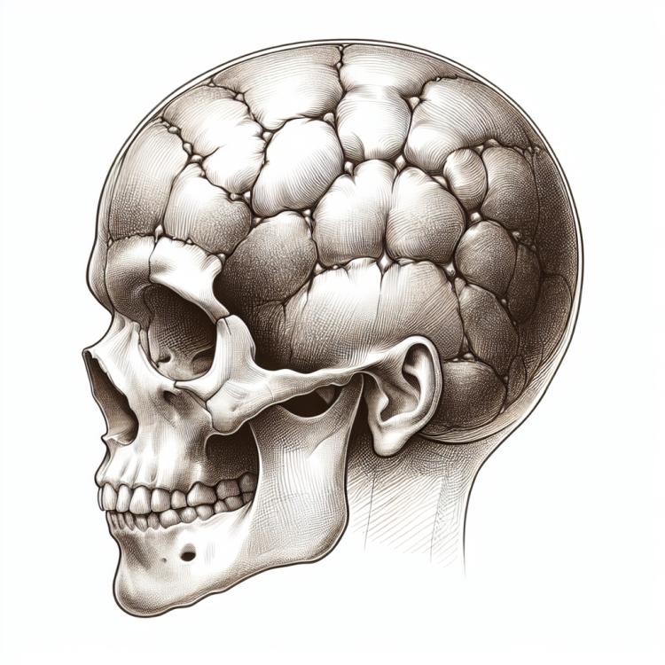
Synostosis is a pathological condition characterized by the premature closure of the sutures in the skull or bone plates, leading to distortion in the growth of the skull and bones. This process can result in severe disruptions in the development and function of the organs and systems of the body. Understanding the essence of synostosis is essential for timely diagnosis and the selection of optimal treatment, as neglecting this condition can have serious consequences for the patient’s health.
Pathophysiology of synostosis
Craniosynostosis is a pathological condition characterized by premature or abnormal fusion of the bones of the skull. The pathophysiology of craniosynostosis is related to a disruption of the normal bone growth activity and the separate development of the skull bones during the period between them. Abnormal fusion of the bones can lead to skull deformities and various neurological complications, such as hydrocephalus and developmental delays in the child. Scientific studies highlight genetic factors, environmental influences, as well as mechanical and biochemical possible causes of craniosynostosis development.
- Premature fusion of bone sutures: the main cause of synostosis lies in the disturbances of the normal growth and separate development of the bones of the skull.
- Gene defects and heredity: genetic factors may contribute to the occurrence of synostosis in children at an early age.
- Environmental impact: factors such as exposure to toxic substances or external environmental influences on fetal development can contribute to the development of synostosis.
- Mechanical injuries: damage to the bones of the skull as a result of injuries, birth trauma, or surgical interventions can lead to synostosis.
- Biochemical disorders: disturbances in the biochemical processes of the body may also affect the development of bones and contribute to the syndrome of premature fusion of the skull bones.
Clinical picture of synostosis
The clinical picture of synostosis can vary depending on the location and degree of fusion of the skull bones. Common symptoms may include abnormalities in head shape, asymmetrical facial features, noticeable concavity or protrusion of the frontal-nasal transition, as well as unusual skull shapes. In children, synostosis can lead to developmental delays, increased pressure in the cranial cavity, and as the bony tissue grows, potential cognitive and neurological consequences. Understanding the clinical picture of synostosis is important for early diagnosis and appropriate treatment to improve patient outcomes.
- Skull shape: deformations of the skull, including unusual protrusions, indentations, or asymmetrical facial features.
- Developmental delay: children with synostosis may experience delays in psychomotor development and general developmental issues.
- Increased intracranial pressure: fusion of bones can lead to increased pressure within the cranial cavity, causing headaches and other symptoms.
- Cognitive impairments: some children with synostosis may experience problems with memory, attention, and other cognitive functions.
- Appearance abnormalities: in addition to head deformities, synostosis can manifest in unusual skull shapes, which may affect the child’s appearance.
Expert opinion: approaches to the treatment of synostosis
The experts’ opinion on approaches to the treatment of synostosis is based on a comprehensive approach that includes conservative and surgical methods. Depending on the type of synostosis, the degree of its progression, and the individual characteristics of the patient, specialists consider options such as observation, the use of orthoses and helmets, as well as surgical interventions to correct the fusion of cranial sutures. Experts believe that timely examination, accurate diagnosis, and adequate treatment can improve the prognosis for patients with synostosis and prevent possible complications in the future.

Methods of diagnosing synostosis
Diagnosis of synostosis includes various examination methods, including clinical examination, X-ray, computed tomography (CT), and magnetic resonance imaging (MRI). Clinical examination allows for the identification of characteristic signs of changes in the shape of the skull and face in the patient. X-ray methods provide additional information about the condition of the skull bones and the head framework, while CT and MRI allow for a more detailed study of the degree and nature of the fusion of the bones.
These diagnostic methods help specialists justify the diagnosis of synostosis, assess its severity and characteristics, which is crucial for choosing the optimal treatment plan. Early and accurate diagnosis of synostosis allows for the necessary therapy to be administered in a timely manner, which can significantly influence the outcome and prognosis for the patient.
- Clinical examination: The doctor should pay attention to changes in the shape of the skull and facial features, which may indicate synostosis.
- X-ray: Radiological examination can detect anomalies and changes in the bony structure of the skull.
- Computed Tomography (CT): CT scanning allows for more detailed images of the skull bones and determination of the degree of fusion of the cranial sutures.
- Magnetic Resonance Imaging (MRI): MRI helps identify structural and functional changes in the brain, which may be important for diagnosing synostosis.
- Consultation with specialists: Specialists in neuroradiology and pediatric neurosurgery may participate in the diagnosis and assessment of synostosis.
Approaches to the treatment of synostosis
-
– Surgical intervention: One of the main treatment methods for synostosis is surgical separation of the fused skull bones in order to restore normal head shape and prevent possible complications.
– Rehabilitation measures: After the operation, patients may require specialized rehabilitation to restore functions of the craniofacial area and to prevent complications in the postoperative period.
– Periodic medical observation: Patients with synostosis syndrome require long-term monitoring by medical professionals to control their condition and the further development of the pathology.
– Individual approach to treatment: Since each case of synostosis is unique, it is important to approach treatment individually, taking into account the patient’s characteristics and the nature of the pathology.
– Consultation with a multidisciplinary team of specialists: In cases of synostosis, consultation with surgeons, neurologists, rehabilitation specialists, and other experts is often required to develop a comprehensive treatment plan.
Prevention measures for synostosis
Additionally, a healthy lifestyle for the pregnant woman, including proper nutrition, abstaining from harmful habits, and taking folic acid, can have a positive impact on the health of the unborn baby and reduce the risk of various anomalies, including synostosis. Overall, prevention of synostosis is based on a comprehensive approach that includes regular health monitoring of the child and support for parents in this process.
- Regular medical check-ups: It is important to have timely visits to specialists for monitoring the child’s development and detecting symptoms of synostosis at early stages.
- Healthy lifestyle during pregnancy: Maintaining a healthy lifestyle during pregnancy, including proper nutrition, avoiding harmful habits, and taking necessary vitamins and minerals.
- Consultation with specialists upon noticing changes: If there are suspicions of any developmental abnormalities, parents should consult doctors for evaluation and further advice.
- Monitoring the child’s development: Parents should regularly track the child’s physical development, paying attention to the shape of the head, skull, and face.
- Adherence to safety measures: Preventing injuries and traumatic head injuries helps reduce the risk of developing synostosis.
Amazing aspects of synostosis
Interesting facts about craniosynostosis include the study of genetic and environmental factors that influence its development, as well as the assessment of the effectiveness of various treatment methods and the impact of late surgical interventions on the prognosis of the condition. Understanding the unique mechanisms underlying craniosynostosis and developing new strategies for diagnosis and treatment are key areas for improving therapy outcomes and the quality of life for patients.