Ureterocele: features of diagnosis and treatment methods
Definition of ureterocele
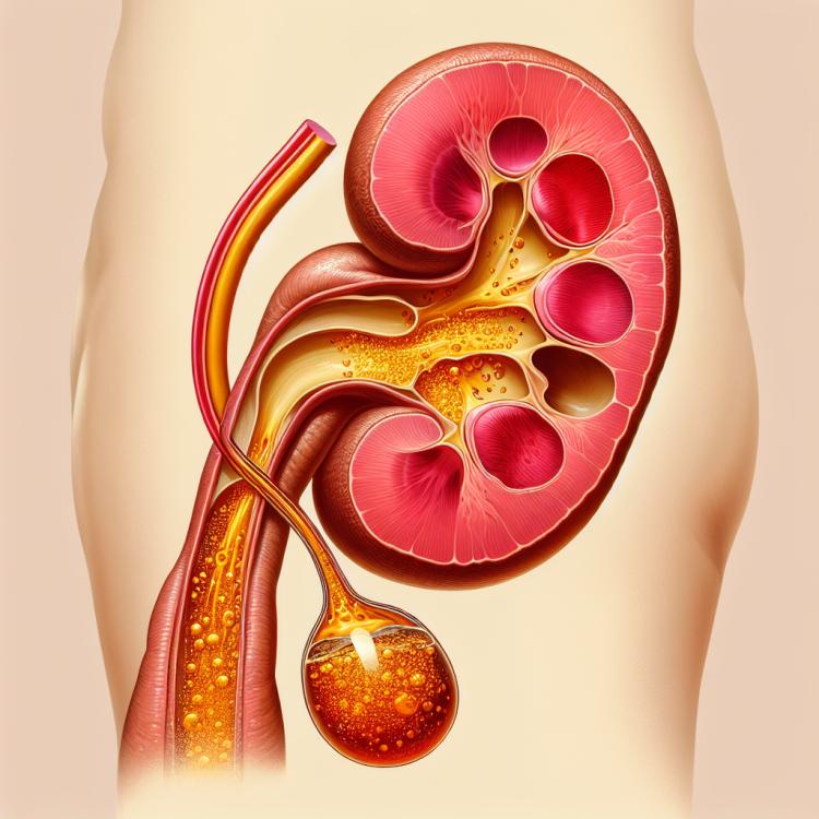
Ureterocele is an expansion of a segment of the ureter, forming a sac-like protrusion in its wall. It is usually caused by abnormal development or a defect in the ureter, which leads to urinary retention and potential obstruction of the urinary tract. Ureterocele can result in symptoms of urinary tract obstruction, such as back pain, frequent urination, or even urinary tract infections. The diagnosis of ureterocele involves various examination methods, such as ultrasound, computed tomography, and cystoscopy, to determine the characteristics and size of the protrusion.
Etiology of ureterocele
Ureterocele is a congenital defect of the ureter, usually caused by incomplete development of the ureteral valve in the fetus. This defect leads to urine retention in the ureter, resulting in distension, which leads to ureterocele. Other causes of ureterocele may include trauma, infections, tumors, or compression of the ureter due to the formation of kidney stones.
- Underdevelopment of the ureteral valve: Incomplete formation of the urethral valve in the fetus becomes the cause of frequent cases of ureterocele.
- Trauma: Injury to the urethra, possibly as a result of surgical interventions or other traumatic events, can contribute to the development of ureterocele.
- Urethral infections: Chronic infections of the urethra can cause inflammation and compression of the urethra, leading to an increased risk of ureterocele.
- Tumor formation: The appearance of tumors in the vicinity of the urethra can lead to compression and pressure on the urethra, promoting the formation of ureterocele.
- Kidney stone formation: Kidney stones can lead to blockage of the urethra, which becomes a risk factor for the development of ureterocele.
Clinical manifestations of ureterocele
The clinical manifestations of ureterocele can vary depending on the size of the ureterocele, the degree of ureteral obstruction, the presence of infection, or associated urological complications. Patients with ureterocele may experience back or abdominal pain, frequent urination, hematuria, elevated blood pressure, as well as symptoms of intoxication.
Other possible symptoms include low levels of vitality, loss of appetite, swelling or edema, and elevated body temperature. The diagnosis of ureterocele is based on clinical manifestations, examination results such as ultrasound or CT scans, as well as urodynamic tests.
- Back and abdominal pain: Patients with ureterocele often complain of back or abdominal pain caused by distension of the ureter.
- Frequent urination: Ureterocele can lead to frequent urges to urinate due to possible obstruction of the ureter.
- Hematuria: The presence of blood in the urine can be one of the signs of ureterocele, caused by the rupture of small vessels in the ureter.
- Increased pressure: Some patients may experience increased pressure as a result of ureteral obstruction and urine retention.
- Symptoms of intoxication: In cases of complications from ureterocele, patients may experience symptoms of intoxication, such as general weakness, nausea, or vomiting.
Approaches to the treatment of ureterocele: expert opinions
Experts in the field of urology typically apply an individual approach to the treatment of ureterocele depending on clinical manifestations, degree of obstruction, and presence of complications. The main goal of treating ureterocele is to ensure normal functioning of the urinary system and to prevent possible complications.
Conservative methods, such as observation, medication therapy, and physiotherapy, may be used in mild cases of ureterocele. However, more serious cases may require surgical intervention, such as endoscopic reconstruction of the ureter or surgical removal of the ureterocele. The decision on the choice of treatment method is usually made collectively by experts, taking into account the individual characteristics of each case.

Methods for diagnosing ureterocele
Various examination methods can be used for the diagnosis of ureterocele, such as ultrasound, computed tomography (CT), and magnetic resonance imaging (MRI). Ultrasound allows visualization of the urinary tract structure and helps determine the size and shape of the ureterocele, as well as assess the presence of urinary tract obstruction. CT and MRI provide a more detailed representation of the anatomy of the urinary system, allowing for a more precise determination of the nature of the ureterocele and potential complications.
Additional diagnostic methods for ureterocele may include radiography (IVP), cystoscopy, and urodynamic tests. The results of these studies help clarify the diagnosis, determine the degree of functional insufficiency of the urinary tract, and choose the most effective treatment method for ureterocele for each specific patient.
- Ultrasound examination: Allows assessment of the structure of the urinary tract and detection of ureterocele.
- Computed tomography (CT): Provides detailed images of the urinary system for accurate determination of the size and characteristics of the ureterocele.
- Magnetic resonance imaging (MRI): Provides information about the structure and function of the ureter and helps determine the features of ureterocele.
- X-ray (IVP): Can be used for additional visualization of the urinary system and confirmation of the ureterocele diagnosis.
- Cystoscopy and urodynamic tests: Allow for a more detailed examination of the urinary tract condition and assessment of the degree of functional insufficiency.
Methods of treating ureterocele
- Conservative treatment: Includes monitoring of the ureterocele, prescribing antibiotics in the presence of infection, painkillers to reduce discomfort, and regular examinations for control.
- Endoscopic procedures: Methods such as cystoscopy and ureteroscopy may be used to repair ureterocele through minimally invasive interventions.
- Surgical reconstruction: Open surgery may be required for large or complex ureteroceles to restore normal anatomy of the urinary tract.
- Robot-assisted surgery: The use of surgical robots to perform precise and complex procedures can be an effective approach in the treatment of ureterocele.
- Pediatric consultation: In children with ureterocele, it is important to conduct treatment under the supervision of a pediatric urologist or pediatric surgeon for optimal results.
Prevention measures for ureterocele
In addition, it is important to avoid traumatic injuries to the urinary tract, adhere to hygiene standards, consume an adequate amount of fluids, maintain a normal level of activity, and lead a healthy lifestyle to reduce the risk of developing ureterocele.
- Regular medical check-up: it is recommended to undergo examination by a urologist for the timely detection of any changes in the urinary tract condition.
- Timely treatment of infections: urological infections should be treated promptly to avoid possible development of ureterocele.
- Genetic counseling: to study possible genetic factors that may influence the development of ureterocele.
- Avoiding traumatic injuries to the urinary tract: take precautions to prevent traumatic injuries, which can lead to ureterocele.
- Healthy lifestyle: maintaining a normal level of activity, proper nutrition, regular physical exercise, and moderate fluid intake helps reduce the risk of developing ureterocele.
Fascinating aspects of ureterocele
Research in the field of ureterocele focuses on improving diagnostic and treatment methods for this condition with the aim of enhancing the prognosis and quality of life for patients. Understanding the mechanisms behind the development of ureterocele and developing innovative therapeutic approaches are of interest to the scientific community and specialists in urology and pediatric surgery.