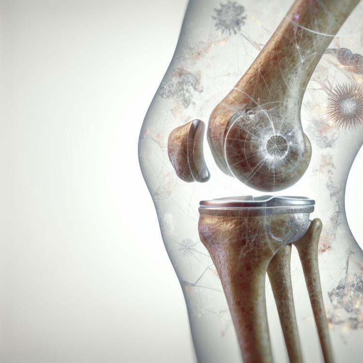
Dislocation of the patella: diagnosis, treatment, and rehabilitation
- Definition and symptoms of patellar dislocation in the section ‘What is Patellar Dislocation’
- Etiology of Patellar Dislocation
- Clinical picture of Patellar Dislocation
- Expert opinion on patellar dislocation therapy
- Methods for diagnosing patellar dislocation
- Treatment strategies for patellar dislocation
- Measures to prevent dislocation of the patella
- Amazing moments around the dislocation of the kneecap
- FAQ
Definition and symptoms of patellar dislocation in the section ‘What is Patellar Dislocation’
A dislocation of the patella is a condition in which the end of the femur comes out of the knee joint. The main symptoms include sudden pain in the knee, a feeling of instability in the joint, swelling, and limited movement.
In a patellar dislocation, there may be a popping or clicking sensation in the joint, as well as swelling of the tissues around the knee. Patients may experience difficulties when walking and standing, and often seek medical attention with complaints of discomfort in the knee area.
Etiology of Patellar Dislocation
Dislocation of the patella is most often caused by traumatic events such as accidents or sports injuries, which can lead to an unnatural position of the knee. Other causes may include congenital anomalies of the knee joint structure or a predisposition to sports injuries due to joint structure characteristics.
Dislocation of the patella can also be caused by excessive pressure on the knee or improper joint movements during physical activity. Factors such as weak muscles or knee instability can increase the risk of this condition.
- Injuries: accidents, sports injuries, and falls can cause patellar dislocation.
- Congenital anomalies: structural features of the knee joint inherited at birth can increase the risk of dislocation.
- Increased pressure: prolonged or excessive pressure on the knee due to improper movements or loads can cause dislocation.
- Sporting activity: intense training or competitions without proper preparation or technique coordination can contribute to the development of patellar dislocation.
- Weak muscles and joint instability: insufficient muscle development around the knee joint or overall joint instability can increase the risk of dislocation.
Clinical picture of Patellar Dislocation
The clinical picture of a patellar dislocation includes characteristic signs such as sharp pain in the knee area, swelling, and limited movement in the joint. The patient may also experience instability and weakness in the knee, which significantly complicates walking and other motor activities.
Additional symptoms may include the appearance of crackling or popping sounds in the joint during movement, bruising and redness at the site of injury, and a possible sensation of the knee “popping out” of the joint. In the case of a patellar dislocation, it is important to conduct a comprehensive examination for an accurate diagnosis and to determine the optimal treatment.
- Sharp Pain: characterized by an acute, intense pain syndrome in the knee area that worsens with movement.
- Swelling and Inflammation: swelling and redness of the injured segment are observed, indicating an inflammatory process.
- Restriction of Movement: a symptom of restricted movement in the knee joint that occurs due to pain or instability when attempting to bend or straighten.
- Knee Instability: the patient may feel uncertainty in the knee, a sense of “shifting,” or an inability to bear weight on the leg due to loss of joint stability.
- Crunching and Clicking: during movement, typical sounds in the joint may be heard, indicating the presence of damage or displacements.
Expert opinion on patellar dislocation therapy
When discussing patellar dislocation therapy, experts highlight the importance of a combined approach to treatment that includes conservative methods, physical therapy, and, in some cases, surgical intervention. Among conservative methods, pain relief medications, physical therapy, and rehabilitation are often used to restore joint function.
Experts emphasize that the correct treatment for patellar dislocation should be individualized and depends on the severity of the injury, the patient’s condition, and the characteristics of their body. Specialists recommend prescribing treatment after a thorough diagnostic examination and consultation with medical experts to achieve optimal results in knee joint recovery.

Methods for diagnosing patellar dislocation
The diagnosis of patellar dislocation includes conducting a physical examination of the joint, as well as using various imaging diagnostic methods, such as X-ray, magnetic resonance imaging (MRI), and computed tomography (CT). These methods allow doctors to obtain a detailed understanding of the condition of the knee joint, assess the structure of the tissues, and determine the presence of dislocation.
Additional diagnostic methods, such as ultrasound examination or arthroscopy, may be used for a more accurate determination of the characteristics of the injury and the selection of the optimal treatment method. Diagnosis is an important stage in patellar dislocation, as the precise determination of the nature of the injury allows for the appointment of effective treatment and the prevention of possible complications.
- Physical examination: The doctor conducts a visual inspection of the knee joint, assessing its structure and functions.
- X-ray: X-ray images are used to assess bone damage and identify joint deformities.
- Magnetic Resonance Imaging (MRI): This imaging method creates detailed images of soft tissues and joint structures, including ligaments and cartilage.
- Computed Tomography (CT): CT allows for three-dimensional images of the knee joint to diagnose various anomalies and injuries.
- Ultrasound examination: Ultrasound can be used to assess soft tissues and ligaments, as well as to identify pathologies in the knee joint.
Treatment strategies for patellar dislocation
Physical therapy, exercises to strengthen the muscles around the knee joint, as well as overall strengthening and stretching of the muscles—all of this can contribute to the recovery of joint functionality after a dislocation. Professional treatment and a rehabilitation plan devised by a specialist play an important role in the recovery process and in preventing recurrences.
- Conservative treatment: Includes physiotherapy, exercises to strengthen muscles, joint stabilization, and the use of special braces.
- Surgical intervention: In complex cases, surgery may be required to restore the stability and functionality of the knee joint.
- Physical rehabilitation: An optimal program of physical therapy and exercises after a dislocation helps to restore the function of the joint.
- Strengthening and stretching muscles: The treatment process includes exercises to strengthen the muscles around the knee and overall strengthening of the muscle corset.
- Professional treatment and rehabilitation: A treatment plan developed by a specialist plays a key role in recovery after a kneecap dislocation.
Measures to prevent dislocation of the patella
In addition, it is important to avoid traumatic situations, use appropriate protection while engaging in sports, and ensure the correct positioning of the joint during physical activities. Regular consultations with specialists, monitoring body weight, and proper nutrition also contribute to overall joint health and reduce the likelihood of kneecap dislocation.
- Regular strength training exercises to strengthen the muscles around the knee joint.
- Stretching exercises and joint flexibility to improve mobility and reduce strain on the knee.
- Avoiding traumatic situations and using protection during sports activities to prevent knee injuries.
- Adhering to the correct technique when performing physical exercises to avoid overloads and injuries.
- Regular consultations with specialists to assess joint condition and develop personalized injury prevention programs.
Amazing moments around the dislocation of the kneecap
Another remarkable aspect is that proper rehabilitation after a patellar dislocation can significantly reduce the risk of future injuries and improve the overall functional state of the joint. Professional and comprehensive treatment, as well as adherence to specialists’ recommendations, play an important role in recovery from this type of injury.